Talk:MitoPedia: Fluorometry: Difference between revisions
From Bioblast
(Undo revision 194027 by Antunes Diana (talk)) |
No edit summary |
||
| (27 intermediate revisions by 3 users not shown) | |||
| Line 1: | Line 1: | ||
{{MitoPedia | |||
|description='''Fluorometry''' (or [[fluorimetry]]) is the general term given to the method of measuring the fluorescent emission of a substance following excitation by light at a shorter wavelength. | |||
}} | |||
__TOC__ | __TOC__ | ||
| Line 19: | Line 21: | ||
== '''O2k-Fluo Smart-Module''' == | == '''O2k-Fluo Smart-Module''' == | ||
=== | === [[O2k-FluoRespirometer]] === | ||
[[ | |||
=== Select the Smart Fluo-Sensors === | === Select the Smart Fluo-Sensors === | ||
| Line 63: | Line 38: | ||
:::: Connect the Smart Fluo-Sensor cable to the Fluo plug of the O2k, insert the male plug of the cable into the female Fluo plug. The red dot on the male plug faces straight upwards. Each Smart Fluo-Sensor can be used on O2k-Chamber A or B. The blue frame of the chamber window and the Smart Fluo-Sensor are specially designed to align in a specific rotational position with the cable extending horizontally to left (chamber A) and right (chamber B). The Smart Fluo-Sensor is carefully inserted into the window frame and rotated into final position, leaving no gap between window frame and sensor body. | :::: Connect the Smart Fluo-Sensor cable to the Fluo plug of the O2k, insert the male plug of the cable into the female Fluo plug. The red dot on the male plug faces straight upwards. Each Smart Fluo-Sensor can be used on O2k-Chamber A or B. The blue frame of the chamber window and the Smart Fluo-Sensor are specially designed to align in a specific rotational position with the cable extending horizontally to left (chamber A) and right (chamber B). The Smart Fluo-Sensor is carefully inserted into the window frame and rotated into final position, leaving no gap between window frame and sensor body. | ||
:::: To remove the Smart Fluo-Sensor, carefully pull out the sensor body with slight back and forth rotations. Do not pull the cable. | :::: To remove the Smart Fluo-Sensor, carefully pull out the sensor body with slight back and forth rotations. Do not pull the cable. | ||
=== Settings in [[DatLab | DatLab 7.4]] === | |||
:::*''-See:'' In the [[O2k control]] window in the [[Amperometric,Amp]] tab, set fluorescence intensity and amplification of the signal. | |||
== '''O2k-Fluo LED2-Module''' == | == '''O2k-Fluo LED2-Module''' == | ||
[[MiPNet17.05 O2k-Fluo LED2-Module]] | [[MiPNet17.05 O2k-Fluo LED2-Module]] | ||
=== Select the Fluorescence-Sensors === | === Select the Fluorescence-Sensors === | ||
:::: Switching between different excitation wavelengths and filters is achieved by simply exchanging the | :::: Switching between different excitation wavelengths and filters is achieved by simply exchanging the Fluo-Sensors. Two types of optical sensors are supplied with different LEDs for fluorescence excitation, and the effective spectra of the LEDs are modified by filters. | ||
:::: Select the Fluo-Sensor and filter set from section 6 of [[MiPNet17.05_O2k-Fluorescence_LED2-Module#Fluorophores|application-specific settings]]. Each Fluo-Sensor is delivered with a mounted filter set. | :::: Select the Fluo-Sensor and filter set from section 6 of [[MiPNet17.05_O2k-Fluorescence_LED2-Module#Fluorophores|application-specific settings]]. Each Fluo-Sensor is delivered with a mounted filter set. | ||
| Line 80: | Line 61: | ||
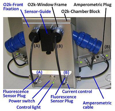
=== Setup and connect O2k-Fluorescence LED2-Module === | === Setup and connect O2k-Fluorescence LED2-Module === | ||
[[File:Fluorescence-Control Unit lettered.jpg|400px|right|link=]] | [[File:Fluorescence-Control Unit lettered.jpg|400px|right|link=]] | ||
::::* Switch off the O2k with the power switch on the rear of the [[O2k-Main Unit]]. | ::::* Switch off the O2k with the power switch on the rear of the [[O2k-Main Unit]]. | ||
::::* Press the power switch on the front panel of the Fluorescence-Control Unit. Check that the two red/green control lights (O2k-Fluorescence LED2-Module B and upwards) or the central green control light are on. | |||
::::* Remove both blue [[O2k-Window Frame]]s. Insert the [[O2k-Window Tool]] around the outer rim of the window frame and unscrew in a counter clockwise direction. | ::::* Remove both blue [[O2k-Window Frame]]s. Insert the [[O2k-Window Tool]] around the outer rim of the window frame and unscrew in a counter clockwise direction. | ||
::::* Remove the '''Sensor-Guide''' (‘nose’) from the '''O2k-Front Fixation''' of the Fluorescence-Control Unit. | ::::* Remove the '''Sensor-Guide''' (‘nose’) from the '''O2k-Front Fixation''' of the Fluorescence-Control Unit. | ||
| Line 89: | Line 71: | ||
:::: In this configuration the O2k can be used for [[high-resolution respirometry]] and fluorometry. It is not necessary to dismount the Fluorescence-Control Unit for basic HRR when a fluorescence signal is not recorded. | :::: In this configuration the O2k can be used for [[high-resolution respirometry]] and fluorometry. It is not necessary to dismount the Fluorescence-Control Unit for basic HRR when a fluorescence signal is not recorded. | ||
=== Settings in DatLab | |||
=== Settings in DatLab: Series E-G === | |||
[[File:Configuration.PNG|right|300px]] | [[File:Configuration.PNG|right|300px]] | ||
::::* From the DatLab menu choose [Oxygraph]/[O2k-Control]. | ::::* From the DatLab menu choose [Oxygraph]/[O2k-Control]. | ||
:::: In the '''O2k configuration | :::: In the '''[[O2k configuration]]''' window include serial number (#) of the sensors used. | ||
:::: In the '''O2k | :::: In the '''[[O2k control]]''' window in the ´Amperometric, Amp´ tab the Fluo/LED intensity and the gain (amplification) can be set. | ||
:::: '''Setting the LED Intensity from DatLab: O2k-Fluorescence LED2-Module Series B and higher (modules <u>without</u> mechanical selector switch)''' | :::: '''Setting the LED Intensity from DatLab: O2k-Fluorescence LED2-Module Series B and higher (modules <u>without</u> mechanical selector switch)''' | ||
::::* Set the desired light intensity (0 to 2000 mv) in the field "Amp Polarization Voltage [mV]". | ::::* Set the desired light intensity (0 to 2000 mv) in the field "Amp Polarization Voltage [mV]". | ||
::::* Click on "Send to Oxygraph" or "Connect to Oxygraph-2k" to apply the new settings. | ::::* Click on "Send to Oxygraph" or "Connect to Oxygraph-2k" to apply the new settings. | ||
::::* If any current >= 0 is set and the Fluorescence Module is switched on, the indicator light below the respective chamber on the Fluorescence-Control Unit will be green. If the current is 0 (the LED is not used) but the Fluorescence-Control_Unit is switched on, the indicator light will be red. | ::::* If any current >= 0 is set and the Fluorescence Module is switched on, the indicator light below the respective chamber on the Fluorescence-Control Unit will be green. If the current is 0 (the LED is not used) but the Fluorescence-Control_Unit is switched on, the indicator light will be red. | ||
:::: Limitations: For O2k-Series D the LED intensity must be set to the same value for both chambers. | :::: Limitations: For O2k-Series D the LED intensity must be set to the same value for both chambers. | ||
Note: The actual current (in mA) used to drive the LED is the value set in DatLab (Amp polarization voltage) divided by 100. | Note: The actual current (in mA) used to drive the LED is the value set in DatLab (Amp polarization voltage) divided by 100. | ||
| Line 126: | Line 105: | ||
|- | |- | ||
|} | |} | ||
== '''General''' == | == '''General''' == | ||
=== Fluorescence dyes === | === Fluorescence dyes === | ||
{| class="wikitable" | {| class="wikitable" | ||
| Line 141: | Line 113: | ||
! Application !!Sensor!! Filter set !! O2k output !! Light intensity (polarization voltage) - ''Note a'' !! Gain !! Comment | ! Application !!Sensor!! Filter set !! O2k output !! Light intensity (polarization voltage) - ''Note a'' !! Gain !! Comment | ||
|- | |- | ||
| [[Amplex UltraRed]]||[[Fluorescence-Sensor Green]] or Smart Fluo-Sensor Green || [[ Filter Set AmR| AmR]] || [[O2k_signal_and_output|Type B]] || 100 - 500 || 1000 (at light intensity = 100) || | | [[Amplex UltraRed]]||[[Fluorescence-Sensor Green]] or Smart Fluo-Sensor Green || [[ Filter Set AmR| AmR]] || [[O2k_signal_and_output|Type B]] || 100 - 500 || 1000 (at light intensity = 100) || at c(AmR) = 10 µM | ||
|- | |- | ||
| [[TMRM]]||[[Fluorescence-Sensor Green]]or Smart Fluo-Sensor Green || [[ Filter Set AmR| AmR]] || [[O2k_signal_and_output|Type C]] || 200 - 500|| 1000 || at c(TMRM) = 2 µM | | [[TMRM]]||[[Fluorescence-Sensor Green]]or Smart Fluo-Sensor Green || [[ Filter Set AmR| AmR]] || [[O2k_signal_and_output|Type C]] || 200 - 500|| 1000 || at c(TMRM) = 2 µM | ||
|- | |- | ||
| [[Safranin]] ||[[Fluorescence-Sensor Blue]]or Smart Fluo-Sensor Blue || [[Filter Set Saf|Saf]] || [[O2k_signal_and_output| Type C]] || 100 - | | [[Safranin]] ||[[Fluorescence-Sensor Blue]]or Smart Fluo-Sensor Blue || [[Filter Set Saf|Saf]] || [[O2k_signal_and_output| Type C]] || 100 - 500 || 1000 || at c(Saf) = 2 µM, | ||
|- | |- | ||
| [[Magnesium green]]||[[Fluorescence-Sensor Blue]]or Smart Fluo-Sensor Blue|| [[Filter Set MgG / CaG| MgG / CaG]] || [[O2k_signal_and_output| Type B]] || 100 - | | [[Magnesium green]]||[[Fluorescence-Sensor Blue]]or Smart Fluo-Sensor Blue|| [[Filter Set MgG / CaG| MgG / CaG]] || [[O2k_signal_and_output| Type B]] || 100 - 500|| 1000 || at c(Mg Green) = 2 µM | ||
|- | |- | ||
| [[Calcium green]]||[[Fluorescence-Sensor Blue]]or Smart Fluo-Sensor Blue|| [[Filter Set MgG / CaG| MgG / CaG]] || [[O2k_signal_and_output|Type A and C]] ||100 -300|| 1000 ||at c(Ca Green) = 2 µM | | [[Calcium green]]||[[Fluorescence-Sensor Blue]]or Smart Fluo-Sensor Blue|| [[Filter Set MgG / CaG| MgG / CaG]] || [[O2k_signal_and_output|Type A and C]] ||100 -300|| 1000 ||at c(Ca Green) = 2 µM | ||
| Line 164: | Line 136: | ||
[[Image:Filter Set AmR.JPG|180px|right]] | [[Image:Filter Set AmR.JPG|180px|right]] | ||
::::* The set of filters for the determination of H<sub>2</sub>O<sub>2</sub> production with [[Amplex UltraRed]] should be used together with [[Fluorescence-Sensor Green | ::::* The set of filters for the determination of H<sub>2</sub>O<sub>2</sub> production with [[Amplex UltraRed]] should be used together with [[Fluorescence-Sensor Green]]. | ||
::::[[File:AmR excitation scan 1mA.png|300px]] | ::::[[File:AmR excitation scan 1mA.png|300px]] | ||
| Line 176: | Line 148: | ||
==== Filter Set Saf ==== | ==== Filter Set Saf ==== | ||
[[Image:Filter_Set_Saf.JPG|180px|right]] | [[Image:Filter_Set_Saf.JPG|180px|right]] | ||
::::* Set of filters for the (qualitative) determination of mitochondrial membrane potential with [[Safranin]] should be used together with [[Fluorescence-Sensor Blue]] or [[Smart Fluo-Sensor Blue | ::::* Set of filters for the (qualitative) determination of mitochondrial membrane potential with [[Safranin]] should be used together with [[Fluorescence-Sensor Blue]] or [[Smart Fluo-Sensor Blue]]. | ||
::::[[File:Saf excitation scan 1mA.png|300px]] | ::::[[File:Saf excitation scan 1mA.png|300px]] | ||
| Line 190: | Line 162: | ||
[[Image:Filter_Set_MgG_CaG.JPG|180px|right]] | [[Image:Filter_Set_MgG_CaG.JPG|180px|right]] | ||
::::* The set of filters for the determination of concentrations of Mg<sup>2+</sup> or Ca<sup>2+</sup> with the fluorophores [[Magnesium green]] and [[Calcium green]], respectively, should be used together with [[Fluorescence-Sensor Blue]] or [[Smart Fluo-Sensor Blue | ::::* The set of filters for the determination of concentrations of Mg<sup>2+</sup> or Ca<sup>2+</sup> with the fluorophores [[Magnesium green]] and [[Calcium green]], respectively, should be used together with [[Fluorescence-Sensor Blue]] or [[Smart Fluo-Sensor Blue]]. | ||
::::[[File:MgG excitation scan 1mA.png|300px]] | ::::[[File:MgG excitation scan 1mA.png|300px]] | ||
| Line 209: | Line 181: | ||
::::The light intensity of the LED ([[Fluorescence-Control_Unit#Control_of_LED-intensity|LED-intensity]]) and the signal amplification ([[Fluorescence-Control_Unit#Gain|Gain]]) can be adjusted in a wide range. The table suggests initial values, which can be optimised for specific applications. | ::::The light intensity of the LED ([[Fluorescence-Control_Unit#Control_of_LED-intensity|LED-intensity]]) and the signal amplification ([[Fluorescence-Control_Unit#Gain|Gain]]) can be adjusted in a wide range. The table suggests initial values, which can be optimised for specific applications. | ||
::::* The settings depend on the concentration of the fluorophore, which vary between different applications. Therefore, only recommendations for specific fluorophore concentrations are given. In the Amplex UltraRed assay the fluorophore is formed during the experiment. | ::::* The settings depend on the concentration of the fluorophore, which vary between different applications. Therefore, only recommendations for specific fluorophore concentrations are given. In the Amplex UltraRed assay the fluorophore is formed during the experiment. | ||
::::* The recommendations apply to experiments at 37 °C. The | ::::* The recommendations apply to experiments at 37 °C. The fluorescence intensity increases strongly at lower temperatures. Then the light intensity is reduced to avoid off-scale signals. | ||
::::* The light intensity of the LEDs is set by the current control, independent for each fluorescence sensor (O2k-Chamber A and B). The current is controlled by DatLab in a very wide range for optimization according to sample and fluorophore requirements. At higher LED-intensity the optical sensitivity is increased, i.e. the signal change per concentration change is enhanced. However, even moderately intensive light may exert negative effects: (i) Damage to the sample reducing the biological activity. (ii) Damage to fluorophores catalyzing degradation and various side reactions. Therefore, the LED-intensity should be kept as low as compatible with a smooth signal, i.e. when the resolution is just not limited by noise or disturbances. The values indicated in the table [[O2k-Fluo_LED2-Module#Application-specific_settings|application specific settings]] are only suggestions to start with. It is recommended to optimize the light intensity specifically for each application. | |||
=== Gain, amplification === | === Gain, amplification === | ||
:::: The gain for the Amp channel can be set in the Control Table of the Oxygraph Control window, the section “Amp” to 1, 10, 100, or 1000. The gain setting will influence the Amp raw signal recorded in Volt. See [[O2k-Fluorescence_LED2-Module#Observing_the_fluorescence_signal| Observing the Fluorescence Signal]]. The amplified signal can be recorded in the range -10 to +10 V. | :::: The gain for the Amp channel can be set in the Control Table of the Oxygraph Control window, the section “Amp” to 1, 10, 100, or 1000. The gain setting will influence the Amp raw signal recorded in Volt. See [[O2k-Fluorescence_LED2-Module#Observing_the_fluorescence_signal| Observing the Fluorescence Signal]]. The amplified signal can be recorded in the range -10 to +10 V. | ||
| Line 272: | Line 247: | ||
::::* [[Titration-Injection microPump |TiP2k]]: Our tests indicated that the fluorometric signal is not affected by the TIP2k needle inserted into the O2k-Chamber. | ::::* [[Titration-Injection microPump |TiP2k]]: Our tests indicated that the fluorometric signal is not affected by the TIP2k needle inserted into the O2k-Chamber. | ||
== '''Troubleshooting''' == | |||
=== Defective Fluo-Sensor or O2k or O2k-LED2 Fluo-Module=== | === Defective Fluo-Sensor or O2k or O2k-LED2 Fluo-Module=== | ||
::: If the raw fluorescence signal is 0 V with and | ::: If the raw fluorescence signal is 0 V with and without fluorescence dyes, it might be possible that your Fluo-Sensor is defective. How can you identify the problem? | ||
:::: Please go through the following check-list: | |||
:::: 1. Check that the [[Illumination on/off|illumination]] is | :::: 1. Check that the [[Illumination on/off|illumination]] is switched off in the ´[[O2k control]]´window. | ||
:::: 2. Check the settings for the Amperometric channel in the ´[[O2k control]]´ window, in the Amperometric,Amp tab. Gain for Fluo-Sensor and the Fluo intensity cannot be 0. | :::: 2. Check the settings for the [[Amperometric,Amp]]channel in the ´[[O2k control]]´ window, in the [[Amperometric,Amp]] tab. Gain for Fluo-Sensor and the Fluo intensity cannot be 0. | ||
:::: 3. The Fluo-Sensors are connected to the O2k and inserted into the O2k-Chamber in the right position (see above in the 1.3 Connect Smart Fluo-Sensor to O2k section). | :::: 3. The Fluo-Sensors are connected to the O2k and inserted into the O2k-Chamber in the right position (see above in the 1.3 Connect Smart Fluo-Sensor to O2k section). | ||
:::: 4. If all the above-mentioned steps are checked and the detected signal with your Fluo-Sensor is 0 | :::: 4. If all the above-mentioned steps are checked and the detected signal with your Fluo-Sensor is 0 (negative or very high) in order to localise the problem, perform the following test. Add distilled water or respiration medium into the O2k-Chamber, insert the black stopper and change the settings for fluorometric measurements (see above): | ||
::::::* 1. run: Fluo-Sensor A in chamber A and Fluo-Sensor B in chamber B | ::::::* 1. run: Fluo-Sensor A in chamber A and Fluo-Sensor B in chamber B | ||
::::::* 2. run: Fluo-Sensor A in chamber B and Fluo-Sensor B in chamber A | ::::::* 2. run: Fluo-Sensor A in chamber B and Fluo-Sensor B in chamber A | ||
:::::: If your Fluo-Sensor A is defective, the fluorescence signal should be 0 V both in chamber A and B. In this case, the Fluo-Sensor should be shipped to us in order to repair it. | :::::: If your Fluo-Sensor A is defective, the fluorescence signal should be 0 V with Fluo-Sensor A both in chamber A and B. In this case, the Fluo-Sensor should be shipped to us in order to repair it. | ||
:::::: If the O2k or the O2k-LED2 | :::::: If the O2k or the O2k-LED2 Fluo-Module are defective, 0 V or negative fluorescence signal can be observed just in one chamber with two different Fluo-Sensors (see example below). In this case, the O2k or the Fluorescence- Control Unit (Series D-G) should be shipped to us in order to repair it. | ||
::::: Please contact our [https://wiki.oroboros.at/index.php/O2k-Open_Support| customer support] to analyse your problem before shippment. Please always send us original DatLab files with the above-mentioned tests to evaluate the defective Fluo-Sensor performance. | ::::: Please contact our [https://wiki.oroboros.at/index.php/O2k-Open_Support| customer support] to analyse your problem before shippment. Please always send us original DatLab files with the above-mentioned tests to evaluate the defective Fluo-Sensor performance. | ||
:::: '''Problem: Defective O2k or O2k-LED2 Fluo-Module''' | :::: '''Problem: Defective O2k or O2k-LED2 Fluo-Module''' | ||
| Line 299: | Line 272: | ||
::::[[File:Hassan Hazirah 2.test.png|700px]] | ::::[[File:Hassan Hazirah 2.test.png|700px]] | ||
::::[[File:Hassan Hazirah biological sample.png|700px]] | ::::[[File:Hassan Hazirah biological sample.png|700px]] | ||
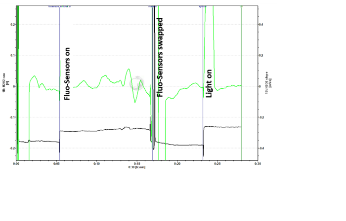
:::: Analysing these tests after changing the Fluo-Sensors between the O2k-Chambers, it is obvious that the problem is related to the O2k or O2k-LED2 | :::: Analysing these tests after changing the Fluo-Sensors between the O2k-Chambers, it is obvious that the problem is related to the O2k or O2k-LED2 Fluo-Module, since the fluorescence signal is negative and unstable in chamber B with both Fluo-Sensors. In the third figure the fluorescence signal is very unstable and it makes measurement unpossible with biological sample. Both the O2k and the LED2 Fluo-Module should be shipped to our electronic workshop. | ||
=== The raw fluorescence signal [V] appears constantly at 10 V === | |||
::: If you see a constant raw signal at 10 V when using the [[O2k-Fluo Smart-Module]] or [[MiPNet17.05 O2k-Fluo LED2-Module | O2k-Fluo LED2-Module]], this is probably due to high light exposure to the photodiode. Check the following parameters: | |||
::::* The chamber illumination must be switched off: Check it in the [[O2k control]] window. | |||
::::* The photodiode filters must be inserted, otherwise the light emitted from the LED will not be filtered. Click [[Filter-Cap#Mounting_a_Filter-Cap|here]] more information on how to mount the filter cap. | |||
::::* Ensure that the [[O2k-Fluo_Smart-Module#Connect_Smart_Fluo-Sensor_to_O2k|Smart Fluo-Sensor is correctly inserted]] at the O2k frame of the chamber window. | |||
::::* Use [[O2k-Fluo_Smart-Module#Stoppers|black PEEK stoppers]]. | |||
=== Negative fluorescence signal=== | === Negative fluorescence signal=== | ||
::::Problem: The fluorescence | ::::Problem: The fluorescence signal of the Fluo-Sensor in chamber B is negative, while the other Fluo-Sensor has ~0 V. The same result occurs when the [https://bioblast.at/index.php/MiPNet17.05_O2k-Fluo_LED2-Module| O2k-LED2 Fluo-Module] is disconnected from the the O2k/switched off. | ||
::::Provided by Anne Laure Charles, FR_Strasbourg_Zoll J,anne laure charles <[email protected] | ::::Provided by Anne Laure Charles, FR_Strasbourg_Zoll J,anne laure charles <[email protected] | ||
::::[[File:Anne Laure Charles files.png|700px]] | ::::[[File:Anne Laure Charles files.png|700px]] | ||
:::: Answer: Connacting our electronic workshop, the problem might be related to wet plug of the | :::: Answer: Connacting our electronic workshop, the problem might be related to a wet plug of the Fluo-Sensors. The solution is to dry it out somehow ''e.g.'' hair dryer and let it run connected to the O2k for a couple of days in dry and warm environment. If these treatments cannot solve the problem and you get still negative fluorescence values, Fluo-Sensor should be sent back to us and our electronic partner will look at it and replace it if needed. | ||
[[Image:BB-Bioblast.jpg|left|40px|link=http://www.bioblast.at/index.php/Bioblast:About|Bioblast wiki]] | [[Image:BB-Bioblast.jpg|left|40px|link=http://www.bioblast.at/index.php/Bioblast:About|Bioblast wiki]] | ||
[[Image:O2k-Publications.jpg|left|116px|link=O2k-Publications: Topics|O2k-Publications in the MiPMap]] | [[Image:O2k-Publications.jpg|left|116px|link=O2k-Publications: Topics|O2k-Publications in the MiPMap]] | ||
| Line 329: | Line 309: | ||
}} | }} | ||
</div> | </div> | ||
== Useful links: == | |||
::::» [[MiPNet22.11 O2k-FluoRespirometer manual| O2k-FluoRespirometer manual]] | |||
::::» [[O2k-Demo_experiments |O2k-Demo experiments]] | |||
::::» [[MiPNet06.05 Specifications | Sole source information]] | |||
::::» [[Oroboros O2k]] | |||
[[Image:SmartFluo.png|thumb|200px|right| Smart Fluo-Sensors]] | |||
[[File:Smart_Fluo-Sensor_-_connect_.png|thumb|100px|right|'''Orientation''' for connection]] | |||
[[File:Smart_Fluo-Sensor_RIGHT.jpg|thumb|100px|right|'''OK''', fully inserted]] | |||
[[File:Smart_Fluo-Sensor_WRONG.jpg|thumb|100px|right|'''WRONG''', not fully inserted]] | |||
[[File:Smart_Fluo-Sensor_inserted.jpg|thumb|100px|right|'''Horizontal''' cable routing]] | |||
=== O2k-Fluorometry Workshops === | |||
::::* [[Oroboros_Events#Next_O2k-Workshops|Oroboros O2k-Workshops]] | |||
== Popular Bioblast page == | == Popular Bioblast page == | ||
Latest revision as of 10:15, 26 May 2020

- high-resolution terminology - matching measurements at high-resolution
Talk:MitoPedia: Fluorometry
Description
Fluorometry (or fluorimetry) is the general term given to the method of measuring the fluorescent emission of a substance following excitation by light at a shorter wavelength.
» MitoPedia methods: Fluorometry
»
MitoPedia O2k and high-resolution respirometry:
O2k hardware

MitoPedia O2k and high-resolution respirometry:
O2k-Open Support
O2k-Fluo Smart-Module
O2k-FluoRespirometer
Select the Smart Fluo-Sensors
- Switching between different excitation wavelengths and filters is achieved by simply exchanging the Smart Fluo-Sensors. Two types of optical sensors are supplied with different LEDs for fluorescence excitation, and the effective spectra of the LEDs are modified by filters.
- Select the Fluo-Sensor and filter set from section 6 of application-specific settings. Each Fluo-Sensor is delivered with a mounted filter set.
Smart Fluo-Sensor Green
- Excitation LED 525 nm (dominant wavelength) with short pass filter, emission (red) with long pass filter, individual sensors are calibrated with sensor-specific memory and direct input into DatLab 7 to obtain reproducible light intensities with different sensors, including photodiode, Filter-Cap equipped with Filter Set AmR for Amplex UltraRed and TMRM measurements when delivered.
- Two units of this item are standard components of the O2k-FluoRespirometer and the O2k-Fluo Smart-Module. Smart Fluo-Sensors can be used with O2k-Series H and higher.
Smart Fluo-Sensor Blue
- Excitation LED 465 nm (dominant wavelenght) with short pass filter, emission (red) with long pass filter, individual sensors are calibrated with sensor-specific memory and direct input into DatLab 7 to obtain reproducible light intensities with different sensors, including photodiode, Filter-Cap equipped with Filter Set Saf for measurement of mitochondrial membrane potential with safranin and rhodamine 123. Filter Set MgG / CaG for Magnesium green and Calcium green measurements are included.
- Two units of this item are standard components of the O2k-FluoRespirometer and the O2k-Fluo Smart-Module. Smart Fluo-Sensors can be used with O2k-Series H and higher.
Setup and connect Smart Fluo-Sensor to O2k
- Connect the Smart Fluo-Sensor cable to the Fluo plug of the O2k, insert the male plug of the cable into the female Fluo plug. The red dot on the male plug faces straight upwards. Each Smart Fluo-Sensor can be used on O2k-Chamber A or B. The blue frame of the chamber window and the Smart Fluo-Sensor are specially designed to align in a specific rotational position with the cable extending horizontally to left (chamber A) and right (chamber B). The Smart Fluo-Sensor is carefully inserted into the window frame and rotated into final position, leaving no gap between window frame and sensor body.
- To remove the Smart Fluo-Sensor, carefully pull out the sensor body with slight back and forth rotations. Do not pull the cable.
Settings in DatLab 7.4
- -See: In the O2k control window in the Amperometric,Amp tab, set fluorescence intensity and amplification of the signal.
O2k-Fluo LED2-Module
MiPNet17.05 O2k-Fluo LED2-Module
Select the Fluorescence-Sensors
- Switching between different excitation wavelengths and filters is achieved by simply exchanging the Fluo-Sensors. Two types of optical sensors are supplied with different LEDs for fluorescence excitation, and the effective spectra of the LEDs are modified by filters.
- Select the Fluo-Sensor and filter set from section 6 of application-specific settings. Each Fluo-Sensor is delivered with a mounted filter set.
Fluorescence-Sensor Green
- Excitation LED 525 nm (dominant wavelength), photodiode, Filter-Cap equipped with Filter Set AmR for Amplex UltraRed measurements when delivered.
- Two units of this item are standard components of the O2k-Fluo LED2-Module.
Fluorescence-Sensor Blue

- Excitation LED 465 nm (dominant wavelength), photodiode, Filter-Cap equipped with Filter Set Saf for measurement of mitochondrial membrane potential with Safranin when delivered. Filter sets for Magnesium green® / Calcium green® measurements are included.
- Two units of this item are standard components of the O2k-Fluo LED2-Module.
Setup and connect O2k-Fluorescence LED2-Module
- Switch off the O2k with the power switch on the rear of the O2k-Main Unit.
- Press the power switch on the front panel of the Fluorescence-Control Unit. Check that the two red/green control lights (O2k-Fluorescence LED2-Module B and upwards) or the central green control light are on.
- Remove both blue O2k-Window Frames. Insert the O2k-Window Tool around the outer rim of the window frame and unscrew in a counter clockwise direction.
- Remove the Sensor-Guide (‘nose’) from the O2k-Front Fixation of the Fluorescence-Control Unit.
- Place the Fluorescence-Control Unit below the O2k-Chamber Block. Align the windows of the O2k-Front-Fixation with the windows of the O2k-Chamber Block, re-insert the O2k-Window Frames, and screw them finger-tight onto the O2k-Main Unit.
- Re-attach the Sensor-Guide to the O2k-Front-Fixation. (There is no need to use the fixation screw supplied with early models).
- Place the power-cables from the rear of the Fluorescence-Control Unit in the middle below the O2k-Main Unit from front to rear. Unplug the mains power cable of the O2k and plug it into the female plug of the Fluorescence-Control Unit. Insert the male plug of the Fluorescence-Control Unit into the mains socket at the rear of the O2k.
- Connect the amperometric cables attached to the side of the Fluorescence-Control Unit to the ‘Amp’ plugs (labeled "NO" in Series D-E) on the O2k-Main Unit.
- In this configuration the O2k can be used for high-resolution respirometry and fluorometry. It is not necessary to dismount the Fluorescence-Control Unit for basic HRR when a fluorescence signal is not recorded.
Settings in DatLab: Series E-G

- From the DatLab menu choose [Oxygraph]/[O2k-Control].
- In the O2k configuration window include serial number (#) of the sensors used.
- In the O2k control window in the ´Amperometric, Amp´ tab the Fluo/LED intensity and the gain (amplification) can be set.
- Setting the LED Intensity from DatLab: O2k-Fluorescence LED2-Module Series B and higher (modules without mechanical selector switch)
- Set the desired light intensity (0 to 2000 mv) in the field "Amp Polarization Voltage [mV]".
- Click on "Send to Oxygraph" or "Connect to Oxygraph-2k" to apply the new settings.
- If any current >= 0 is set and the Fluorescence Module is switched on, the indicator light below the respective chamber on the Fluorescence-Control Unit will be green. If the current is 0 (the LED is not used) but the Fluorescence-Control_Unit is switched on, the indicator light will be red.
- Limitations: For O2k-Series D the LED intensity must be set to the same value for both chambers.
- Setting the LED Intensity from DatLab: O2k-Fluorescence LED2-Module Series B and higher (modules without mechanical selector switch)
Note: The actual current (in mA) used to drive the LED is the value set in DatLab (Amp polarization voltage) divided by 100.
Previously we quoted the suggested current. To simplify  the operation we now state directly the required DatLab settings.
- Setting the LED Intensity from DatLab: O2k-Fluorescence LED2-Module Series A (modules with mechanical selector switch)
- O2k-Series A of the O2k-Fluorescence LED2-Module has a mechanical selector switch for each chamber on the front side of the Fluorescence-Control_Unit
- Set the switch located on the front panel of the Fluorescence-Control_Unit to position 9 with help of the small screw driver included with the Module.
- Follow the procedure described above for Series B and higher.
- For LED2-Module Series A it is still possible to set the LED current directly at the Fluorescence-Control_Unit. However, such set function is no longer in use. The relevant instructions can be found on the discussion page in the archive.
- Conversion of the LED intensities between the Fluorescence-Sensor of the O2k-Fluo LED2-Module (O2k-Series D to G) and the Smart Fluo-Sensor of the O2k-FluoRespirometer (O2k-Series H)
- The O2k-FluoRespirometer (O2k-Series H) and software DatLab 7, the fluorescent intensity (excitation) of the Smart Fluo-Sensors is set in 'Fluo intensity' units. The Smart Fluo-Sensors are pre-calibrated to guarantee similar fluorescence intensity among Smart Fluo-Sensors of the same colour, storing the sensor-specific 'gain value' in an internal memory. Technically speaking, DatLab 7 (i) reads this 'gain value' from a connected Smart Fluo-Sensor to adjust the output intensity and (ii) automatically corrects for the non-linear relationship between intensity and 'mV' to output the correct fluorescent intensity. In contrast, the fluorescent intensity (excitation) of the Fluorescence-Sensors of the O2k-Fluo LED2-Module (O2k-Series D to G) is set directly in 'mV' units. To set the same intensity for O2k-Fluo LED2-Module (O2k-Series D to G) Fluorescence-Sensor as for a O2k-FluoRespirometer (O2k-Series H) Smart Fluo-Sensor, we have created an Excel sheet that allows to calculate it.
General
Fluorescence dyes
| Application | Sensor | Filter set | O2k output | Light intensity (polarization voltage) - Note a | Gain | Comment |
|---|---|---|---|---|---|---|
| Amplex UltraRed | Fluorescence-Sensor Green or Smart Fluo-Sensor Green | AmR | Type B | 100 - 500 | 1000 (at light intensity = 100) | at c(AmR) = 10 µM |
| TMRM | Fluorescence-Sensor Greenor Smart Fluo-Sensor Green | AmR | Type C | 200 - 500 | 1000 | at c(TMRM) = 2 µM |
| Safranin | Fluorescence-Sensor Blueor Smart Fluo-Sensor Blue | Saf | Type C | 100 - 500 | 1000 | at c(Saf) = 2 µM, |
| Magnesium green | Fluorescence-Sensor Blueor Smart Fluo-Sensor Blue | MgG / CaG | Type B | 100 - 500 | 1000 | at c(Mg Green) = 2 µM |
| Calcium green | Fluorescence-Sensor Blueor Smart Fluo-Sensor Blue | MgG / CaG | Type A and C | 100 -300 | 1000 | at c(Ca Green) = 2 µM |
| Rhodamine 123 | Fluorescence-Sensor Blueor Smart Fluo-Sensor Blue | Filter Set Saf | Type III | 100 -300 | 1000 | at c(Rh123) = 1 µM |
Stoppers
- Use only black PEEK stoppers in conjunction with fluorometric measurements. The black stoppers can be used for all HRR applications. See MiPNet22.11 O2k-FluoRespirometer manual for calibration of the O2k-chamber volume, which is identical for PEEK and PVDF stoppers.
Selecting a Filter Set
- The Filter-Cap of each sensor can be removed for application of various filter combinations on the same optical sensor.
- Selection of a Filter Set: >> Application specific settings.
- The Filter-Cap of each sensor can be removed for application of various filter combinations on the same optical sensor.
Filter Set AmR

- The set of filters for the determination of H2O2 production with Amplex UltraRed should be used together with Fluorescence-Sensor Green.

- Excitation spectrum obtained with the Fluorescence-Sensor Green at 1 mA current supply and equipped with the Filter Set AmR.
- In the O2k-Fluo LED2-Module dye based-absorption filters in form of polymer films are used. If required, cut-off values related to the filters (short of a full spectrum) can be used:
- AmR excitation filter (SG 370): dye based short pass filter, 50% T: 535 nm, suppressing (<5% Transmission) lambda > 595 nm
- AmR emission filter (RL2001): dye based long pass filter, 50% T: 620 nm, suppressing (<2% transmission) lambda < 590 nm
- The spectra of excitation (LED filter: R370) and emission (photodiode filter: R2001) can be found on the webpage of our supplier, ROSCO: https://emea.rosco.com/de/node/1706 (please select the filter numbers to download the specific data sheets).
Filter Set Saf

- Set of filters for the (qualitative) determination of mitochondrial membrane potential with Safranin should be used together with Fluorescence-Sensor Blue or Smart Fluo-Sensor Blue.

- Excitation spectrum obtained with the Fluorescence-Sensor Blue at 1mA current supply and equipped with the Filter Set Saf.
- In the O2k-Fluo LED2-Module dye based absorption filters in form of polymer films are used. If required, cut-off values related to the filters (short of a full spectrum) can be used:
- Saf excitation filter: (SG357): dye based short pass filter, 50% T: 470 nm, suppressing (<5% Transmission) lambda > 530 nm
- Saf emission filter: (SG19): dye based long pass filter, 50% T: 600 nm, suppressing (<1% transmission) lambda < 550 nm
- The spectra of excitation (LED filter: R357) and emission (photodiode filter: R19) can be found on the webpage of our supplier, ROSCO: https://emea.rosco.com/de/node/1706 (please select the filter numbers to download the specific data sheets).
Filter Set MgG/CaG

- The set of filters for the determination of concentrations of Mg2+ or Ca2+ with the fluorophores Magnesium green and Calcium green, respectively, should be used together with Fluorescence-Sensor Blue or Smart Fluo-Sensor Blue.

- Excitation spectrum obtained with the Fluorescence-Sensor Blue at 1 mA current supply and equipped with the filter set for Magnesium green® / Calcium green®.
- In the O2k-Fluo LED2-Module dye based absorption filters in form of polymer films are used. If required, cut-off values related to the filters (short of a full spectrum) can be used:
- MgG/CaG excitation filter (SG59):dye based short pass filter, 50% T: not applicable (max. transmission < 50%), suppressing (<5% Transmission) lambda > 490 nm
- MgG/CaG emission filter (RL21):dye based long pass filter,50% T: 570 nm, suppressing (<1% transmission) lambda < 525 nm
- The spectra of excitation (LED filter: R59) and emission (photodiode filter: R21) can be found on the webpage of our supplier, ROSCO: https://emea.rosco.com/de/node/1706 (please select the filter numbers to download the specific data sheets).
Mounting a Filter-Cap
- Applies to: O2k-Fluo LED2-Module (O2k-Series D to G) Fluorescence Sensor and O2k-FluoRespirometer (O2k-Series H) with Smart Fluo-Sensors
- Dismounting: Pull the Filter-Cap straight from the sensor. The Filter-Cap Guide prevents rotational movements.
- Remove all filters and store them in the filter box labeled for this Filter Set.
- Insert the filters form the selected Filter Set: The round filters of each Filter Set fit to the round window of the Filter-Cap and cover the LED, the rectangular filters fit into the rectangular window of the Filter-Cap and cover the photodiode.
- Mounting: Hold sensor and filter cap in a vertical position above you. Align the Filter-Cap with the Filter-Cap Guide (small steel rod) protruding from the sensor. Press the Filter-Cap onto the sensor without rotational movements.
Control of LED-intensity
- The light intensity of the LED (LED-intensity) and the signal amplification (Gain) can be adjusted in a wide range. The table suggests initial values, which can be optimised for specific applications.
- The settings depend on the concentration of the fluorophore, which vary between different applications. Therefore, only recommendations for specific fluorophore concentrations are given. In the Amplex UltraRed assay the fluorophore is formed during the experiment.
- The recommendations apply to experiments at 37 °C. The fluorescence intensity increases strongly at lower temperatures. Then the light intensity is reduced to avoid off-scale signals.
- The light intensity of the LED (LED-intensity) and the signal amplification (Gain) can be adjusted in a wide range. The table suggests initial values, which can be optimised for specific applications.
- The light intensity of the LEDs is set by the current control, independent for each fluorescence sensor (O2k-Chamber A and B). The current is controlled by DatLab in a very wide range for optimization according to sample and fluorophore requirements. At higher LED-intensity the optical sensitivity is increased, i.e. the signal change per concentration change is enhanced. However, even moderately intensive light may exert negative effects: (i) Damage to the sample reducing the biological activity. (ii) Damage to fluorophores catalyzing degradation and various side reactions. Therefore, the LED-intensity should be kept as low as compatible with a smooth signal, i.e. when the resolution is just not limited by noise or disturbances. The values indicated in the table application specific settings are only suggestions to start with. It is recommended to optimize the light intensity specifically for each application.
Gain, amplification
- The gain for the Amp channel can be set in the Control Table of the Oxygraph Control window, the section “Amp” to 1, 10, 100, or 1000. The gain setting will influence the Amp raw signal recorded in Volt. See Observing the Fluorescence Signal. The amplified signal can be recorded in the range -10 to +10 V.
- The gain setting should be chosen to obtain a maximum signal well below 10 V. If the maximum observed raw signal was 9 V in an initial experiment, then the gain should be reduced to avoid "off scale" (>9.99 V). On the other hand, if the maximum recorded raw signal was considerable lower than 1 V (e.g. 0.2 V), the gain can be increased to avoid limitation of resolution by digital noise.
- Note for advanced users: At gain 1, a current of 1 nA is recorded as a voltage of 1 mV (0.001 V). At gain 100, 1 nA corresponds to 100 mV (0.1 V), as can be seen form the table below:
1
|
0.001 | 1000
|
+-10000
|
333
|
10
|
0.01 | 100
|
+-1000
|
33
|
1000
|
1 | 1
|
+-10
|
0.3
|
The fluorescence signal
- O2k signal: The O2k-Fluo LED2-Module is operated through the amperometric (Amp)-Channel of the O2k, with electric current (ampere [Amp]) as the primary signal.
- O2k output: type A, B, or C, or combinations.

Graph layout: Three plots are available in DatLab based on the recorded signal: Amp Raw Signal, Amp Calibrated, and Amp Slope. These plots can be selected from the drop-down lines and displayed with their check boxes either on the Y1 or Y2 [Graph layout / Select Plots].
- Amp Raw Signal displays the raw voltage (including amplification) as recorded by the O2k at a given gain setting.
- Amp Calibrated is the signal after calibration with the parameters set in the O2k-MultiSensor Calibration window.
- Amp slope is the time derivative of the calibrated signal, multiplied by 1000, in units [mV(conc. Unit during calibration)/s], so if the signal was calibrated in µM [nmol/ml] the unit of the slope is pmol/(s ml). To obtain the slope of the raw signal check the appropriate box in the calibration window (DatLab 5.1.0.130 and above).
- Graphs can be generated to display oxygen and fluorescence data, or several graphs can be added to display oxygen and fluorescence data separately. Layout templates are provided, which can be modified and saved as appropriate. All graph settings can be saved as user-defined layouts, see MiPNet19.01C DatLab Guide.
O2k-Fluorometry and the TIP2k
- TiP2k: Our tests indicated that the fluorometric signal is not affected by the TIP2k needle inserted into the O2k-Chamber.
Troubleshooting
Defective Fluo-Sensor or O2k or O2k-LED2 Fluo-Module
- If the raw fluorescence signal is 0 V with and without fluorescence dyes, it might be possible that your Fluo-Sensor is defective. How can you identify the problem?
- Please go through the following check-list:
- 1. Check that the illumination is switched off in the ´O2k control´window.
- If the raw fluorescence signal is 0 V with and without fluorescence dyes, it might be possible that your Fluo-Sensor is defective. How can you identify the problem?
- 2. Check the settings for the Amperometric,Ampchannel in the ´O2k control´ window, in the Amperometric,Amp tab. Gain for Fluo-Sensor and the Fluo intensity cannot be 0.
- 3. The Fluo-Sensors are connected to the O2k and inserted into the O2k-Chamber in the right position (see above in the 1.3 Connect Smart Fluo-Sensor to O2k section).
- 4. If all the above-mentioned steps are checked and the detected signal with your Fluo-Sensor is 0 (negative or very high) in order to localise the problem, perform the following test. Add distilled water or respiration medium into the O2k-Chamber, insert the black stopper and change the settings for fluorometric measurements (see above):
- 1. run: Fluo-Sensor A in chamber A and Fluo-Sensor B in chamber B
- 2. run: Fluo-Sensor A in chamber B and Fluo-Sensor B in chamber A
- If your Fluo-Sensor A is defective, the fluorescence signal should be 0 V with Fluo-Sensor A both in chamber A and B. In this case, the Fluo-Sensor should be shipped to us in order to repair it.
- If the O2k or the O2k-LED2 Fluo-Module are defective, 0 V or negative fluorescence signal can be observed just in one chamber with two different Fluo-Sensors (see example below). In this case, the O2k or the Fluorescence- Control Unit (Series D-G) should be shipped to us in order to repair it.
- Please contact our customer support to analyse your problem before shippment. Please always send us original DatLab files with the above-mentioned tests to evaluate the defective Fluo-Sensor performance.
- Problem: Defective O2k or O2k-LED2 Fluo-Module
- Provided by Hazirah Hassan, University Kebangsaan Malaysia, [email protected]
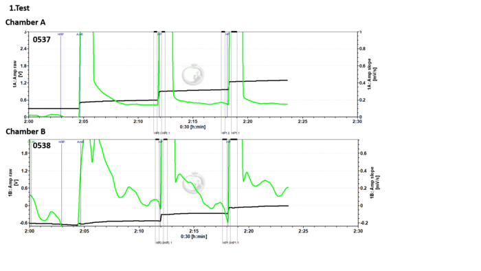
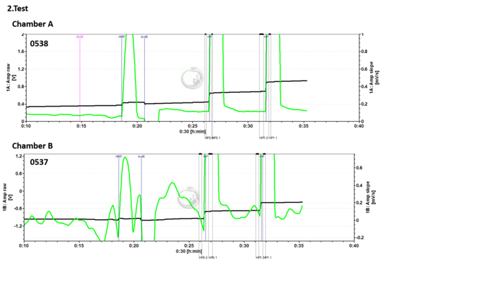
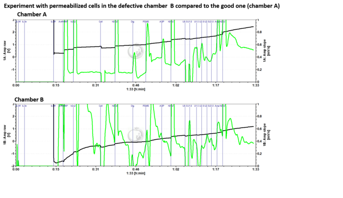
- Analysing these tests after changing the Fluo-Sensors between the O2k-Chambers, it is obvious that the problem is related to the O2k or O2k-LED2 Fluo-Module, since the fluorescence signal is negative and unstable in chamber B with both Fluo-Sensors. In the third figure the fluorescence signal is very unstable and it makes measurement unpossible with biological sample. Both the O2k and the LED2 Fluo-Module should be shipped to our electronic workshop.
- 4. If all the above-mentioned steps are checked and the detected signal with your Fluo-Sensor is 0 (negative or very high) in order to localise the problem, perform the following test. Add distilled water or respiration medium into the O2k-Chamber, insert the black stopper and change the settings for fluorometric measurements (see above):


The raw fluorescence signal [V] appears constantly at 10 V
- If you see a constant raw signal at 10 V when using the O2k-Fluo Smart-Module or O2k-Fluo LED2-Module, this is probably due to high light exposure to the photodiode. Check the following parameters:
- The chamber illumination must be switched off: Check it in the O2k control window.
- The photodiode filters must be inserted, otherwise the light emitted from the LED will not be filtered. Click here more information on how to mount the filter cap.
- Ensure that the Smart Fluo-Sensor is correctly inserted at the O2k frame of the chamber window.
- Use black PEEK stoppers.
- If you see a constant raw signal at 10 V when using the O2k-Fluo Smart-Module or O2k-Fluo LED2-Module, this is probably due to high light exposure to the photodiode. Check the following parameters:
Negative fluorescence signal
- Problem: The fluorescence signal of the Fluo-Sensor in chamber B is negative, while the other Fluo-Sensor has ~0 V. The same result occurs when the O2k-LED2 Fluo-Module is disconnected from the the O2k/switched off.
- Provided by Anne Laure Charles, FR_Strasbourg_Zoll J,anne laure charles <[email protected]

- Answer: Connacting our electronic workshop, the problem might be related to a wet plug of the Fluo-Sensors. The solution is to dry it out somehow e.g. hair dryer and let it run connected to the O2k for a couple of days in dry and warm environment. If these treatments cannot solve the problem and you get still negative fluorescence values, Fluo-Sensor should be sent back to us and our electronic partner will look at it and replace it if needed.

- » References
Sort in ascending/descending order by a click on one of the small symbols in squares below.
Default sorting: chronological. Empty fields appear first in ascending order.
| Was published in year | Has title | Organism | Tissue;cell | |
|---|---|---|---|---|
| MiPNet18.10 O2k-Specifications | 2024-03-26 | Human | Fibroblast | |
| Lhuissier 2024 iScience | 2024 | Lhuissier C, Desquiret-Dumas V, Girona A, Alban J, Faure J, Cassereau J, Codron P, Lenaers G, Baris OR, Gueguen N, Chevrollier A (2024) Mitochondrial F0F1-ATP synthase governs the induction of mitochondrial fission. iScience 27:109808. https://doi.org/10.1016/j.isci.2024.109808 | Mouse | Fibroblast |
| Balmaceda 2024 Biochim Biophys Acta Mol Basis Dis | 2024 | Balmaceda V, Komlodi T, Szibor M, Gnaiger E, Moore AL, Fernandez-Vizarra E, Viscomi C (2024) The striking differences in the bioenergetics of brain and liver mitochondria are enhanced in mitochondrial disease. Biochim Biophys Acta Mol Basis Dis 1870:167033. https://doi.org/10.1016/j.bbadis.2024.167033 | Mouse | Nervous system Liver |
| Ravasz 2024 Sci Rep | 2024 | Ravasz D, Bui D, Nazarian S, Pallag G, Karnok N, Roberts J, Marzullo BP, Tennant DA, Greenwood B, Kitayev A, Hill C, Komlódi T, Doerrier C, Cunatova K, Fernandez-Vizarra E, Gnaiger E, Kiebish Michael A, Raska A, Kolev K, Czumbel B, Narain NR, Seyfried TN, Chinopoulos C (2024) Residual Complex I activity and amphidirectional Complex II operation support glutamate catabolism through mtSLP in anoxia. Sci Rep 14:1729. https://doi.org/10.1038/s41598-024-51365-4 | Mouse | Heart Liver |
| Fitzgerald 2024 J Cachexia Sarcopenia Muscle | 2024 | Fitzgerald LF, Lackey J, Moussa A, Shah SV, Castellanos AM, Khan S, Schonk M, Thome T, Salyers ZR, Jakkidi N, Kim K, Yang Q, Hepple RT, Ryan TE (2024) Chronic aryl hydrocarbon receptor activity impairs muscle mitochondrial function with tobacco smoking. https://doi.org/10.1002/jcsm.13439 | Mouse | Skeletal muscle |
| Al-Sabri 2024 Sci Rep | 2024 | Al-Sabri MH, Ammar N, Korzh S, Alsehli AM, Hosseini K, Fredriksson R, Mwinyi J, Williams MJ, Boukhatmi H, Schiöth HB (2024) Fluvastatin-induced myofibrillar damage is associated with elevated ROS, and impaired fatty acid oxidation, and is preceded by mitochondrial morphological changes. https://doi.org/10.1038/s41598-024-53446-w | Drosophila | Skeletal muscle |
| Donnelly 2024 Redox Biol | 2024 | Donnelly C, Komlódi T, Cecatto C, Cardoso LHD, Compagnion A-C, Matera A, Tavernari D, Campiche O, Paolicelli RC, Zanou N, Kayser B, Gnaiger E, Place N (2024) Functional hypoxia reduces mitochondrial calcium uptake. Redox Biol 71:103037. https://doi.org/10.1016/j.redox.2024.103037 | Human Mouse | Heart Skeletal muscle |
| Cefis 2024 Acta Physiol (Oxf) | 2024 | Cefis M, Dargegen M, Marcangeli V, Taherkhani S, Dulac M, Leduc-Gaudet JP, Mayaki D, Hussain SNA, Gouspillou G (2024) MFN2 overexpression in skeletal muscles of young and old mice causes a mild hypertrophy without altering mitochondrial respiration and H2O2 emission. Acta Physiol (Oxf) [Epub ahead of print]. https://doi.org/10.1111/apha.14119 | Mouse | Skeletal muscle |
| Taylor 2024 Pilot Feasibility Stud | 2024 | Taylor MK, Burns JM, Choi IY, Herda TJ, Lee P, Smith AN, Sullivan DK, Swerdlow RH, Wilkins HM (2024) Protocol for a single-arm, pilot trial of creatine monohydrate supplementation in patients with Alzheimer's disease. Pilot Feasibility Stud 10:42. https://doi.org/10.1186/s40814-024-01469-5 | Human | Lymphocyte Platelet |
| Meldau 2024 Mol Genet Metab Rep | 2024 | Meldau S, Ackermann S, Riordan G, van der Watt GF, Spencer C, Raga S, Khan K, Blackhurst DM, van der Westhuizen FH (2024) A novel mitochondrial DNA variant in MT-ND6: m.14430A>C p.(Trp82Gly) identified in a patient with Leigh syndrome and complex I deficiency. Mol Genet Metab Rep 39:101078. https://doi.org/10.1016/j.ymgmr.2024.101078 | Human | Fibroblast |
| Sorby-Adams 2024 Redox Biol | 2024 | Sorby-Adams A, Prime TA, Miljkovic JL, Prag HA, Krieg T, Murphy MP (2024) A model of mitochondrial superoxide production during ischaemia-reperfusion injury for therapeutic development and mechanistic understanding. Redox Biol 72:103161. https://doi.org/10.1016/j.redox.2024.103161 | Rat | Heart |
| Dominguez-Lopez 2023 Neuropharmacology | 2023 | Dominguez-Lopez S, Ahn B, Sataranatarajan K, Ranjit R, Premkumar P, Van Remmen H, Beckstead MJ (2023) Long-term methamphetamine self-administration increases mesolimbic mitochondrial oxygen consumption and decreases striatal glutathione. https://doi.org/10.1016/j.neuropharm.2023.109436 | Mouse | Nervous system |
| Mahmoud 2023 Pharmacol Res | 2023 | Mahmoud AM, Kostrzewa M, Marolda V, Cerasuolo M, Maccarinelli F, Coltrini D, Rezzola S, Giacomini A, Mollica MP, Motta A, Paris D, Zorzano A, Marzo VD, Ronca R, Ligresti A (2023) Cannabidiol alters mitochondrial bioenergetics via VDAC1 and triggers cell death in hormone-refractory prostate cancer. https://doi.org/10.1016/j.phrs.2023.106683 | Mouse | Endothelial;epithelial;mesothelial cell |
| Gautam 2023 Neurobiol Dis | 2023 | Gautam M, Genç B, Helmold B, Ahrens A, Kuka J, Makrecka-Kuka M, Günay A, Koçak N, Aguilar-Wickings IR, Keefe D, Zheng G, Swaminathan S, Redmon M, Zariwala HA, Özdinler PH (2023) SBT-272 improves TDP-43 pathology in ALS upper motor neurons by modulating mitochondrial integrity, motility, and function. https://doi.org/10.1016/j.nbd.2023.106022 | Rat | Heart Nervous system |
| Donnelly 2023 MitoFit | 2023 | Donnelly C, Komlódi T, Cecatto C, Cardoso LHD, Compagnion AC, Matera A, Tavernari D, Zanou N, Kayser B, Gnaiger E, Place N (2023) Functional hypoxia reduces mitochondrial calcium uptake. MitoFit Preprints 2023.2. https://doi.org/10.26124/mitofit:2023-0002 — 2024-11-17 published in Redox Biol. | Human Mouse | Skeletal muscle Heart Nervous system Other cell lines |
| Leduc-Gaudet 2023 Nat Commun | 2023 | Leduc-Gaudet JP, Franco-Romero A, Cefis M, Moamer A, Broering FE, Milan G, Sartori R, Chaffer TJ, Dulac M, Marcangeli V, Mayaki D, Huck L, Shams A, Morais JA, Duchesne E, Lochmuller H, Sandri M, Hussain SNA, Gouspillou G (2023) MYTHO is a novel regulator of skeletal muscle autophagy and integrity. https://doi.org/10.1038/s41467-023-36817-1 | Mouse | Skeletal muscle |
| Koizumi 2023 Front Cardiovasc Med | 2023 | Koizumi T, Watanabe M, Yokota T, Tsuda M, Handa H, Koya J, Nishino K, Tatsuta D, Natsui H, Kadosaka T, Koya T, Nakao M, Hagiwara H, Kamada R, Temma T, Tanaka S, Anzai T (2023) Empagliflozin suppresses mitochondrial reactive oxygen species generation and mitigates the inducibility of atrial fibrillation in diabetic rats. Front Cardiovasc Med 10: 1005408. | Rat | Heart |
| Salmon 2023 Geroscience | 2023 | Salmón P, Millet C, Selman C, Monaghan P, Dawson NJ (2023) Tissue-specific reductions in mitochondrial efficiency and increased ROS release rates during ageing in zebra finches, Taeniopygia guttata. https://doi.org/10.1007/s11357-022-00624-1 | Birds | Skeletal muscle Liver |
| Devaux 2023 J Comp Physiol B | 2023 | Devaux JBL, Hedges CP, Birch N, Herbert N, Renshaw GMC, Hickey AJR (2023) Electron transfer and ROS production in brain mitochondria of intertidal and subtidal triplefin fish (Tripterygiidae). https://doi.org/10.1007/s00360-023-01495-4 | Fishes | Nervous system |
| Czyzowska 2023 Redox Biol | 2023 | Czyżowska A, Brown J, Xu H, Sataranatarajan K, Kinter M, Tyrell VJ, O'Donnell VB, Van Remmen H (2023) Elevated phospholipid hydroperoxide glutathione peroxidase (GPX4) expression modulates oxylipin formation and inhibits age-related skeletal muscle atrophy and weakness. https://doi.org/10.1016/j.redox.2023.102761 | Mouse | Skeletal muscle |
| Som 2023 Am J Physiol Cell Physiol | 2023 | Som R, Fink BD, Yu L, Sivitz WI (2023) Oxaloacetate regulates complex II respiration in brown fat: dependence on UCP1 expression. Am J Physiol Cell Physiol 324:C1236-48. doi: 10.1152/ajpcell.00565.2022 | Mouse | Fat |
| Steffen 2023 J Exp Biol | 2023 | Steffen JBM, Sokolov EP, Bock C, Sokolova IM (2023) Combined effects of salinity and intermittent hypoxia on mitochondrial capacity and reactive oxygen species efflux in the Pacific oyster, Crassostrea gigas. https://doi.org/10.1242/jeb.246164 | Molluscs | Lung;gill |
| Fletcher 2023 Transl Res | 2023 | Fletcher E, Miserlis D, Sorokolet K, Wilburn D, Bradley C, Papoutsi E, Wilkinson T, Ring A, Ferrer L, Haynatzki G, Smith RS, Bohannon WT, Koutakis P (2023) Diet-induced obesity augments ischemic myopathy and functional decline in a murine model of peripheral artery disease. https://doi.org/10.1016/j.trsl.2023.05.002 | Mouse | Skeletal muscle |
| Pharaoh 2023 Geroscience | 2023 | Pharaoh G, Kamat V, Kannan S, Stuppard RS, Whitson J, Martín-Pérez M, Qian WJ, MacCoss MJ, Villén J, Rabinovitch P, Campbell MD, Sweet IR, Marcinek DJ (2023) The mitochondrially targeted peptide elamipretide (SS-31) improves ADP sensitivity in aged mitochondria by increasing uptake through the adenine nucleotide translocator (ANT). https://doi.org/10.1007/s11357-023-00861-y | Mouse | Skeletal muscle |
| Krause 2023 J Transl Med | 2023 | Krause J, Nickel A, Madsen A, Aitken-Buck HM, Stoter AMS, Schrapers J, Ojeda F, Geiger K, Kern M, Kohlhaas M, Bertero E, Hofmockel P, Hübner F, Assum I, Heinig M, Müller C, Hansen A, Krause T, Park DD, Just S, Aïssi D, Börnigen D, Lindner D, Friedrich N, Alhussini K, Bening C, Schnabel RB, Karakas M, Iacoviello L, Salomaa V, Linneberg A, Tunstall-Pedoe H, Kuulasmaa K, Kirchhof P, Blankenberg S, Christ T, Eschenhagen T, Lamberts RR, Maack C, Stenzig J, Zeller T (2023) An arrhythmogenic metabolite in atrial fibrillation. https://doi.org/10.1186/s12967-023-04420-z | Human | Heart |
| Zvejniece 2023 Biomed Pharmacother | 2023 | Zvejniece L, Svalbe B, Vavers E, Ozola M, Grinberga S, Gukalova B, Sevostjanovs E, Liepinsh E, Dambrova M (2023) Decreased long-chain acylcarnitine content increases mitochondrial coupling efficiency and prevents ischemia-induced brain damage in rats. | Rat | |
| Batterson 2023 Physiol Rep | 2023 | Batterson PM, McGowan EM, Borowik AK, Kinter MT, Miller BF, Newsom SA, Robinson MM (2023) High-fat diet increases electron transfer flavoprotein synthesis and lipid respiration in skeletal muscle during exercise training in female mice. https://doi.org/10.14814/phy2.15840 | Mouse | Skeletal muscle |
| Stouth 2023 Autophagy | 2023 | Stouth DW, vanLieshout TL, Mikhail AI, Ng SY, Raziee R, Edgett BA, Vasam G, Webb EK, Gilotra KS, Markou M, Pineda HC, Bettencourt-Mora BG, Noor H, Moll Z, Bittner ME, Gurd BJ, Menzies KJ, Ljubicic V (2023) CARM1 drives mitophagy and autophagy flux during fasting-induced skeletal muscle atrophy. https://doi.org/10.1080/15548627.2023.2288528 | Mouse | Skeletal muscle |
| Dabrowska 2023 Int J Mol Sci | 2023 | Dabrowska A, Zajac M, Bednarczyk P, Lukasiak A (2023) Effect of quercetin on mitoBKCa channel and mitochondrial function in human bronchial epithelial cells exposed to particulate matter. Int J Mol Sci 24:638. https://doi.org/10.3390/ijms24010638 | Human | Lung;gill Endothelial;epithelial;mesothelial cell |
| Davis 2023 Am J Physiol Regul Integr Comp Physiol . | 2023 | Davis MS, Bayly WM, Hansen CM, Barrett MR, Blake CA (2023) Effects of hyperthermia and acidosis on mitochondrial production of reactive oxygen species. Am J Physiol Regul Integr Comp Physiol . 325(6):R725-R734. | Horse | Skeletal muscle |
| Komlodi 2022 BEC | 2022 | Komlódi T, Tretter L (2022) The protonmotive force – not merely membrane potential. Bioenerg Commun 2022.16. https://doi.org/10.26124/bec:2022-0016 | ||
| Pileggi 2022 EBioMedicine | 2022 | Pileggi CA, Blondin DP, Hooks BG, Parmar G, Alecu I, Patten DA, Cuillerier A, O'Dwyer C, Thrush AB, Fullerton MD, Bennett SAL, Doucet É, Haman F, Cuperlovic-Culf M, McPherson R, Dent RRM, Harper ME (2022) Exercise training enhances muscle mitochondrial metabolism in diet-resistant obesity. https://doi.org/10.1016/j.ebiom.2022.104192 | Human | Skeletal muscle |
| Munro 2022 Mitochondrion | 2022 | Munro D, Rodríguez E, Blier PU (2022) The longest-lived metazoan, Arctica islandica, exhibits high mitochondrial H2O2 removal capacities. https://doi.org/10.1016/j.mito.2022.11.005 | Molluscs | Lung;gill Endothelial;epithelial;mesothelial cell |
| Quemeneur 2022 Sci Rep | 2022 | Quéméneur JB, Danion M, Cabon J, Collet S, Zambonino-Infante JL, Salin K (2022) The relationships between growth rate and mitochondrial metabolism varies over time. https://doi.org/10.1038/s41598-022-20428-9 | Fishes | Skeletal muscle |
| Ceja-Galicia 2022 Antioxidants (Basel) | 2022 | Ceja-Galicia ZA, García-Arroyo FE, Aparicio-Trejo OE, El-Hafidi M, Gonzaga-Sánchez G, León-Contreras JC, Hernández-Pando R, Guevara-Cruz M, Tovar AR, Rojas-Morales P, Aranda-Rivera AK, Sánchez-Lozada LG, Tapia E, Pedraza-Chaverri J (2022) Therapeutic effect of curcumin on 5/6Nx hypertriglyceridemia: association with the improvement of renal mitochondrial β-oxidation and lipid metabolism in kidney and liver. https://doi.org/10.3390/antiox11112195 | Rat | Kidney |
| Brown 2022 Redox Biol | 2022 | Brown JL, Peelor FF 3rd, Georgescu C, Wren JD, Kinter M, Tyrrell VJ, O'Donnell VB, Miller BF, Van Remmen H (2022) Lipid hydroperoxides and oxylipins are mediators of denervation induced muscle atrophy. https://doi.org/10.1016/j.redox.2022.102518 | Mouse | Skeletal muscle |
| Xu 2022 Sci Adv | 2022 | Xu H, Ahn B, Van Remmen H (2022) Impact of aging and oxidative stress on specific components of excitation contraction coupling in regulating force generation. https://doi.org/10.1126/sciadv.add7377 | Mouse | Skeletal muscle |
| Formiga-Jr 2022 J Vis Exp | 2022 | Formiga-Jr MA, Camacho-Pereira J (2022) Assessing mitochondrial function in sciatic nerve by high-resolution respirometry. https://doi.org/10.3791/63690 | Mouse | Nervous system |
| Sarabhai 2022 Diabetologia | 2022 | Sarabhai T, Mastrototaro L, Kahl S, Bönhof GJ, Jonuscheit M, Bobrov P, Katsuyama H, Guthoff R, Wolkersdorfer M, Herder C, Meuth SG, Dreyer S, Roden M (2022) Hyperbaric oxygen rapidly improves tissue-specific insulin sensitivity and mitochondrial capacity in humans with type 2 diabetes: a randomised placebo-controlled crossover trial. https://doi.org/10.1007/s00125-022-05797-0 | Human | Skeletal muscle Fat |
| McKenna 2022 J Appl Physiol (1985) | 2022 | McKenna CF, Salvador AF, Keeble AR, Khan NA, De Lisio M, Konopka AR, Paluska SA, Burd NA (2022) Muscle strength after resistance training correlates to mediators of muscle mass and mitochondrial respiration in middle-aged adults. https://doi.org/10.1152/japplphysiol.00186.2022 | Human | Skeletal muscle |
| Belosludtseva 2022 Membranes (Basel) | 2022 | Belosludtseva NV, Pavlik LL, Belosludtsev KN, Saris NL, Shigaeva MI, Mironova GD (2022) The short-term opening of cyclosporin A-independent palmitate/Sr 2+-induced pore can underlie ion efflux in the oscillatory mode of functioning of rat liver mitochondria. https://doi.org/10.3390/membranes12070667 | Rat | Liver |
| VanLieshout 2022 Mol Metab | 2022 | vanLieshout TL, Stouth DW, Hartel NG, Vasam G, Ng SY, Webb EK, Rebalka IA, Mikhail AI, Graham NA, Menzies KJ, Hawke TJ, Ljubicic V (2022) The CARM1 transcriptome and arginine methylproteome mediate skeletal muscle integrative biology. https://doi.org/10.1016/j.molmet.2022.101555 | Mouse | Skeletal muscle |
| Smith 2022 J Pineal Res | 2022 | Smith KLM, Swiderska A, Lock MC, Graham L, Iswari W, Choudhary T, Thomas D, Kowash HM, Desforges M, Cottrell EC, Trafford AW, Giussani DA, Galli GLJ (2022) Chronic developmental hypoxia alters mitochondrial oxidative capacity and reactive oxygen species production in the fetal rat heart in a sex-dependent manner. https://doi.org/10.1111/jpi.12821 | Rat | Heart |
| Jelenik 2022 Pharmacol Res | 2022 | Jelenik T, Kodde A, Pesta D, Phielix E, Oosting A, Rohbeck E, Dewidar B, Mastrototaro L, Trenkamp S, Keijer J, van der Beek EM, Roden M (2022) Dietary lipid droplet structure in postnatal life improves hepatic energy and lipid metabolism in a mouse model for postnatal programming. https://doi.org/10.1016/j.phrs.2022.106193 | Mouse | Skeletal muscle Liver |
| Salyers 2022 JVS Vasc Sci | 2022 | Salyers ZR, Mariani V, Balestrieri N, Kumar RA, Vugman NA, Thome T, Villani KR, Berceli SA, Scali ST, Vasilakos G, Ryan TE (2022) S100A8 and S100A9 are elevated in chronically threatened ischemic limb muscle and induce ischemic mitochondrial pathology in mice. https://doi.org/10.1016/j.jvssci.2022.03.003 | Human Mouse | Skeletal muscle |
| Starr 2022 Curr Res Physiol | 2022 | Starr VJ, Dzialowski EM (2022) Developing chicken cardiac muscle mitochondria are resistant to variations in incubation oxygen levels. https://doi.org/10.1016/j.crphys.2022.03.001 | Chicken | Heart |
| Loughland 2022 J Exp Biol | 2022 | Loughland I, Lau GY, Jolly J, Seebacher F (2022) Rates of warming impact oxidative stress in zebrafish (Danio rerio). | Zebrafish | Skeletal muscle |
| Yoval-Sanchez 2022 Redox Biol | 2022 | Yoval-Sánchez B, Ansari F, James J, Niatsetskaya Z, Sosunov S, Filipenko P, Tikhonova IG, Ten V, Wittig I, Rafikov R, Galkin A (2022) Redox-dependent loss of flavin by mitochondria complex I is different in brain and heart. https://doi.org/10.1016/j.redox.2022.102258 | Mouse | Heart Nervous system |
| Lopes 2022 Int J Mol Sci | 2022 | Lopes JA, Collino F, Rodrigues-Ferreira C, Sampaio LDS, Costa-Sarmento G, Wendt CHC, Almeida FP, Miranda KR, Kasai-Brunswick TH, Lindoso RS, Vieyra A (2022) Early effects of extracellular vesicles secreted by adipose tissue mesenchymal cells in renal ischemia followed by reperfusion: mechanisms rely on a decrease in mitochondrial anion superoxide production. https://doi.org/10.3390/ijms23062906 | Human | Kidney |
| Qvit 2022 Pharmaceuticals (Basel) | 2022 | Qvit N, Lin AJ, Elezaby A, Ostberg NP, Campos JC, Ferreira JCB, Mochly-Rosen D (2022) A selective inhibitor of cardiac troponin I phosphorylation by delta protein kinase C (δPKC) as a treatment for ischemia-reperfusion injury. https://doi.org/10.3390/ph15030271 | Rat | Heart |
| Hansen 2022 Free Radic Biol Med | 2022 | Hansen C, Møller S, Ehlers T, Wickham KA, Bangsbo J, Gliemann L, Hellsten Y (2022) Redox balance in human skeletal muscle-derived endothelial cells - Effect of exercise training. https://doi.org/10.1016/j.freeradbiomed.2021.12.265 | Human | Endothelial;epithelial;mesothelial cell |
| Yamauchi 2022 Sci Rep | 2022 | Yamauchi Y, Nakamura A, Yokota T, Takahashi K, Kawata S, Tsuchida K, Omori K, Nomoto H, Kameda H, Cho KY, Anzai T, Tanaka S, Terauchi Y, Miyoshi H, Atsumi T (2022) Luseogliflozin preserves the pancreatic beta-cell mass and function in db/db mice by improving mitochondrial function. Sci Rep 12: 9740. | Mouse | Islet cell;pancreas;thymus |
| Pallag 2022 Int J Mol Sci | 2022 | Pallag G, Nazarian S, Ravasz D, Bui D, Komlódi T, Doerrier C, Gnaiger E, Seyfried TN, Chinopoulos C (2022) Proline oxidation supports mitochondrial ATP production when Complex I is inhibited. https://doi.org/10.3390/ijms23095111 | Mouse | Liver Kidney |
| Cruz-Gregorio 2022 Antioxidants (Basel) | 2022 | Cruz-Gregorio A, Aranda-Rivera AK, Aparicio-Trejo OE, Medina-Campos ON, Sciutto E, Fragoso G, Pedraza-Chaverri J (2022) GK-1 Induces oxidative stress, mitochondrial dysfunction, decreased membrane potential, and impaired autophagy flux in a mouse model of breast cancer. https://doi.org/10.3390/antiox12010056 | Mouse | Endothelial;epithelial;mesothelial cell |
| Vandenberg 2022 Am J Physiol Cell Physiol | 2022 | Vandenberg GG, Thotakura A, Scott AL (2022) Mitochondrial bioenergetics of astrocytes in Fragile X syndrome: new perspectives on culture conditions and sex effects. Am J Physiol Cell Physiol 322:C125-35. https://doi.org/10.1152/ajpcell.00130.2021 | Mouse | Nervous system |
| Barath 2022 Int J Mol Sci | 2022 | Baráth B, Jász DK, Horváth T, Baráth B, Maróti G, Strifler G, Varga G, Sándor L, Perényi D, Tallósy S, Donka T, Jávor P, Boros M, Hartmann P (2022) Mitochondrial side effects of surgical prophylactic antibiotics Ceftriaxone and Rifaximin lead to bowel mucosal damage. Int J Mol Sci 23:5064. https://doi.org/10.3390/ijms23095064 | Rat | Liver |
| MiPNet24.10 H2O2 flux analysis | 2021-10-22 |  | ||
| MiPNet26.10 MgG data analysis | 2021-09-16 | | ||
| MiPNet17.05 O2k-Fluo LED2-Module | 2021-06-22 |  | ||
| Jasz 2021 J Cell Mol Med | 2021 | Jász DK, Szilágyi ÁL, Tuboly E, Baráth B, Márton AR, Varga P, Varga G, Érces D, Mohácsi Á, Szabó A, Bozó R, Gömöri K, Görbe A, Boros M, Hartmann P (2021) Reduction in hypoxia-reoxygenation-induced myocardial mitochondrial damage with exogenous methane. https://doi.org/10.1111/jcmm.16498 | Rat | Heart |
| Elliehausen 2021 Exp Gerontol | 2021 | Elliehausen CJ, Minton DM, Nichol AD, Konopka AR (2021) Skeletal muscle mitochondrial respiration in a model of age-related osteoarthritis is impaired after dietary rapamycin. https://doi.org/10.1016/j.exger.2021.111579 | Guinea pig | Skeletal muscle |
| Pharaoh 2021 JCSM Rapid Commun | 2021 | Pharaoh G, Brown J, Ranjit R, Ungvari Z, Van Remmen H (2021) Reduced adenosine diphosphate sensitivity in skeletal muscle mitochondria increases reactive oxygen species production in mouse models of aging and oxidative stress but not denervation. https://doi.org/10.1002/rco2.29 | Mouse | Skeletal muscle |
| Christiansen 2021 Sci Rep | 2021 | Christiansen LB, Dohlmann TL, Ludvigsen TP, Parfieniuk E, Ciborowski M, Szczerbinski L, Kretowski A, Desler C, Tiano L, Orlando P, Martinussen T, Olsen LH, Larsen S (2021) Atorvastatin impairs liver mitochondrial function in obese Göttingen Minipigs but heart and skeletal muscle are not affected. Sci Rep 11:2167. | Pig | Heart Skeletal muscle Liver |
| Yardeni 2021 Proc Natl Acad Sci U S A | 2021 | Yardeni T, Cristancho AG, McCoy AJ, Schaefer PM, McManus MJ, Marsh ED, Wallace DC (2021) An mtDNA mutant mouse demonstrates that mitochondrial deficiency can result in autism endophenotypes. Proc Natl Acad Sci U S A 118:e2021429118. | Mouse | Nervous system |
| Thoral 2021 Biol Lett | 2021 | Thoral E, Roussel D, Chinopoulos C, Teulier L, Salin K (2021) Low oxygen levels can help to prevent the detrimental effect of acute warming on mitochondrial efficiency in fish. Biol Lett 17:20200759. | Fishes | Skeletal muscle |
| Furihata 2021 Commun Biol | 2021 | Furihata T, Takada S, Kakutani N, Maekawa S, Tsuda M, Matsumoto J, Mizushima W, Fukushima A, Yokota T, Enzan N, Matsushima S, Handa H, Fumoto Y, Nio-Kobayashi J, Iwanaga T, Tanaka S, Tsutsui H, Sabe H, Kinugawa S (2021) Cardiac-specific loss of mitoNEET expression is linked with age-related heart failure. Commun Biol 4:138. | Mouse | Heart |
| Salin 2021 Mar Environ Res | 2021 | Salin K, Mathieu-Resuge M, Graziano N, Dubillot E, Le Grand F, Soudant P, Vagner M (2021) The relationship between membrane fatty acid content and mitochondrial efficiency differs within- and between- omega-3 dietary treatments. Mar Environ Res 163:105205. | Fishes | Skeletal muscle |
| Furihata 2021 BMC Pharmacol Toxicol | 2021 | Furihata T, Maekawa S, Takada S, Kakutani N, Nambu H, Shirakawa R, Yokota T, Kinugawa S (2021) Premedication with pioglitazone prevents doxorubicin-induced left ventricular dysfunction in mice. BMC Pharmacol Toxicol 22:27. | Mouse | Heart |
| Cardoso 2021 BEC MgG | 2021 | Cardoso LHD, Doerrier C, Gnaiger E (2021) Magnesium Green for fluorometric measurement of ATP production does not interfere with mitochondrial respiration. Bioenerg Commun 2021.1. https://doi.org/10.26124/bec:2021-0001 | Mouse | Heart |
| Deline 2021 Biochim Biophys Acta Bioenerg | 2021 | Deline ML, Grashei M, van Heijster FHA, Schilling F, Straub J, Fromme T (2021) Adenylate kinase derived ATP shapes respiration and calcium storage of isolated mitochondria. Biochim Biophys Acta Bioenerg 1862:148409. | Mouse | Liver |
| Morgan 2021 Resuscitation | 2021 | Morgan RW, Sutton RM, Himebauch AS, Roberts AL, Landis WP, Lin Y, Starr J, Ranganathan A, Delso N, Mavroudis CD, Volk L, Slovis J, Marquez AM, Nadkarni VM, Hefti M, Berg RA, Kilbaugh TJ (2021) A randomized and blinded trial of inhaled nitric oxide in a piglet model of pediatric cardiopulmonary resuscitation. Resuscitation 162:274-83 . | Pig | Nervous system |
| Cheng 2021 Comp Biochem Physiol B Biochem Mol Biol | 2021 | Cheng H, Munro D, Huynh K, Pamenter ME (2021) Naked mole-rat skeletal muscle mitochondria exhibit minimal functional plasticity in acute or chronic hypoxia. Comp Biochem Physiol B Biochem Mol Biol 255:110596. | Other mammals | Skeletal muscle |
| Vandenberg 2021 Neurochem Int | 2021 | Vandenberg GG, Dawson NJ, Head A, Scott GR, Scott AL (2021) Astrocyte-mediated disruption of ROS homeostasis in Fragile X mouse model. Neurochem Int 146:105036. | Mouse | Nervous system |
| Hansen 2021 Physiol Rep | 2021 | Hansen C, Olsen K, Pilegaard H, Bangsbo J, Gliemann L, Hellsten Y (2021) High metabolic substrate load induces mitochondrial dysfunction in rat skeletal muscle microvascular endothelial cells. Physiol Rep 9:e14855. | Rat | Skeletal muscle Endothelial;epithelial;mesothelial cell |
| Newsom 2021 Med Sci Sports Exerc | 2021 | Newsom SA, Stierwalt HD, Ehrlicher SE, Robinson MM (2021) Substrate-specific respiration of isolated skeletal muscle mitochondria after 1 h of moderate cycling in sedentary adults. https://doi.org/10.1249/mss.0000000000002615 | Human | Skeletal muscle |
| Marrocco 2021 J Immunol | 2021 | Marrocco A, Frawley K, Pearce LL, Peterson J, O'Brien JP, Mullett SJ, Wendell SG, St Croix CM, Mischler SE, Ortiz LA (2021) Metabolic adaptation of macrophages as mechanism of defense against crystalline silica. J Immunol 207:1627-40. | Mouse | Macrophage-derived |
| Apostolopoulou 2021 Sci Adv | 2021 | Apostolopoulou M, Mastrototaro L, Hartwig S, Pesta D, Straßburger K, de Filippo E, Jelenik T, Karusheva Y, Gancheva S, Markgraf D, Herder C, Nair KS, Reichert AS, Lehr S, Müssig K, Al-Hasani H, Szendroedi J, Roden M (2021) Metabolic responsiveness to training depends on insulin sensitivity and protein content of exosomes in insulin-resistant males. https://doi.org/10.1126/sciadv.abi9551 | Human | Skeletal muscle |
| Dall 2021 J Biol Chem | 2021 | Dall M, Hassing AS, Niu L, Nielsen TS, Ingerslev LR, Sulek K, Trammell SAJ, Gillum MP, Barrès R, Larsen S, Poulsen SS, Mann M, Ørskov C, Treebak JT (2021) Hepatocyte-specific perturbation of NAD+ biosynthetic pathways in mice induces reversible nonalcoholic steatohepatitis-like phenotypes. J Biol Chem 297:101388. | Mouse | Liver |
| Riguet 2021 Nat Commun | 2021 | Riguet N, Mahul-Mellier AL, Maharjan N, Burtscher J, Croisier M, Knott G, Hastings J, Patin A, Reiterer V, Farhan H, Nasarov S, Lashuel HA (2021) Nuclear and cytoplasmic huntingtin inclusions exhibit distinct biochemical composition, interactome and ultrastructural properties. Nat Commun 12:6579. | Human | HEK |
| Sarabhai 2021 Diabetologia | 2021 | Sarabhai T, Koliaki C, Mastrototaro L, Kahl S, Pesta D, Apostolopoulou M, Wolkersdorfer M, Bönner AC, Bobrov P, Markgraf DF, Herder C, Roden M (2021) Dietary palmitate and oleate differently modulate insulin sensitivity in human skeletal muscle. Diabetologia 65:301-14. | Human | Skeletal muscle |
| Theall 2021 Physiol Rep | 2021 | Theall B, Stampley J, Cho E, Granger J, Johannsen NM, Irving BA, Spielmann G (2021) Impact of acute exercise on peripheral blood mononuclear cells nutrient sensing and mitochondrial oxidative capacity in healthy young adults. Physiol Rep 9:e15147. | Human | Blood cells |
| Komlodi 2021 BEC AmR-O2 | 2021 | Komlódi T, Sobotka O, Gnaiger E (2021) Facts and artefacts on the oxygen dependence of hydrogen peroxide production using Amplex UltraRed. Bioenerg Commun 2021.4. https://doi.org/10.26124/bec:2021-0004 | Saccharomyces cerevisiae | Other cell lines |
| Galli 2021 Front Physiol | 2021 | Galli GLJ, Ruhr IM, Crossley J, Crossley DA 2nd (2021) The long-term effects of developmental hypoxia on cardiac mitochondrial function in snapping turtles. Front Physiol 12:689684. doi: 10.3389/fphys.2021.689684 | Reptiles | Heart |
| MiPNet24.08 Safranin Analysis Template | 2020-05-13 | | ||
| MiPNet25.14 TPP Analysis Template | 2020-##-## | | ||
| Smith 2020 J Biol Chem | 2020 | Smith CD, Schmidt CA, Lin CT, Fisher-Wellman KH, Neufer PD (2020) Flux through mitochondrial redox circuits linked to nicotinamide nucleotide transhydrogenase generates counterbalance changes in energy expenditure. J Biol Chem 295:16207-16. | Mouse | Skeletal muscle |
| Zussman 2020 J Autoimmun | 2020 | Zussman R, Xu LY, Damani T, Groom KM, Chen Q, Seers B, Viall CA, Chamley LW, Hickey A (2020) Antiphospholipid antibodies can specifically target placental mitochondria and induce ROS production. J Autoimmun 111:102437. | Human | Genital |
| Li Puma 2020 Am J Physiol Regul Integr Comp Physiol | 2020 | Li Puma LC, Hedges M, Heckman JM, Mathias AB, Engstrom MR, Brown AB, Chicco AJ (2020) Experimental oxygen concentration influences rates of mitochondrial hydrogen peroxide release from cardiac and skeletal muscle preparations. Am J Physiol Regul Integr Comp Physiol 318:972-80. | Mouse | Heart Skeletal muscle |
| Fuller 2020 J Appl Physiol (1985) | 2020 | Fuller KNZ, McCoin CS, Allen J, Bell-Glenn S, Koestler DC, Dorn Ii GW, Thyfault JP (2020) Sex and BNIP3 genotype, rather than acute lipid injection, modulate hepatic mitochondrial function and steatosis risk in mice. J Appl Physiol (1985) 128:1251-61. | Mouse | Liver |
| Hartmann 2020 Free Radic Res | 2020 | Hartmann DD, Gonçalves DF, Da Rosa PC, Martins RP, Courtes AA, Franco JL, A Soares FA, Puntel GO (2020) A single muscle contusion promotes an immediate alteration in mitochondrial bioenergetics response in skeletal muscle fibres with different metabolism. Free Radic Res 54:137-49. | Rat | Skeletal muscle |
| Hassan 2020 MitoFit Preprint Arch | 2020 | Hassan Hazirah, Gnaiger Erich, Zakaria Fazaine, Makpol Suzana, Abdul Karim Norwahidah (2020) Alterations in mitochondrial respiratory capacity and membrane potential: a link between mitochondrial dysregulation and autism. https://doi.org/10.26124/mitofit:200003 | Human | |
| Aparicio-Trejo 2020 Free Radic Biol Med | 2020 | Aparicio-Trejo OE, Avila-Rojas SH, Tapia E, Rojas-Morales P, León-Contreras JC, Martínez-Klimova E, Hernández-Pando R, Sánchez-Lozada LG, Pedraza-Chaverri J (2020) Chronic impairment of mitochondrial bioenergetics and β-oxidation promotes experimental AKI-to-CKD transition induced by folic acid. Free Radic Biol Med 154:18-32. | Rat | Kidney |
| Buch 2020 FASEB J | 2020 | Buch BT, Halling JF, Ringholm S, Gudiksen A, Kjøbsted R, Olsen MA, Wojtaszewski JFP, Pilegaard H (2020) Colchicine treatment impairs skeletal muscle mitochondrial function and insulin sensitivity in an age-specific manner. FASEB J 34:8653-70. | Mouse | Skeletal muscle |
| Lozano 2020 Part Fibre Toxicol | 2020 | Lozano O, Silva-Platas C, Chapoy-Villanueva H, Pérez BE, Lees JG, Ramachandra CJA, Contreras-Torres FF, Lázaro-Alfaro A, Luna-Figueroa E, Bernal-Ramírez J, Gordillo-Galeano A, Benitez A, Oropeza-Almazán Y, Castillo EC, Koh PL, Hausenloy DJ, Lim SY, García-Rivas G (2020) Amorphous SiO2 nanoparticles promote cardiac dysfunction via the opening of the mitochondrial permeability transition pore in rat heart and human cardiomyocytes. Part Fibre Toxicol 17:15. | Rat Human | Heart |
| Chausse 2020 Brain Behav Immun | 2020 | Chausse B, Lewen A, Poschet G, Kann O (2020) Selective inhibition of mitochondrial respiratory complexes controls the transition of microglia into a neurotoxic phenotype in situ. Brain Behav Immun 88:802-14. | Mouse | Nervous system |
| Brunetta 2020 J Physiol | 2020 | Brunetta HS, Politis-Barber V, Petrick HL, Dennis KMJH, Kirsh AJ, Barbeau PA, Nunes EA, Holloway GP (2020) Nitrate attenuates HFD-induced glucose intolerance in association with reduced epididymal adipose tissue inflammation and mitochondrial ROS emission. J Physiol 598:3357-71. | Mouse | Fat |
| Ponce 2020 J Am Heart Assoc | 2020 | Ponce JM, Coen G, Spitler KM, Dragisic N, Martins I, Hinton A Jr, Mungai M, Tadinada SM, Zhang H, Oudit GY, Song LS, Li N, Sicinski P, Strack S, Abel ED, Mitchell C, Hall DD, Grueter CE (2020) Stress-induced cyclin C translocation regulates cardiac mitochondrial dynamics. J Am Heart Assoc 9:e014366. | Rat Mouse | Heart |
| Stepanova 2020 Methods Cell Biol | 2020 | Stepanova A, Galkin A (2020) Measurement of mitochondrial H2O2 production under varying O2 tensions. https://doi.org/10.1016/bs.mcb.2019.12.008 | Mouse | Nervous system |
| Mendham 2020 Sci Rep | 2020 | Mendham Amy E, Larsen Steen, George Cindy, Adams Kevin, Hauksson Jon, Olsson Tommy, Fortuin-de Smidt Melony C, Nono Nankam Pamela A, Hakim Olah, Goff Louise M, Pheiffer Carmen, Goedecke Julia H (2020) Exercise training results in depot-specific adaptations to adipose tissue mitochondrial function. Sci Rep 10:3785. | Fat | |
| Hafstad 2020 Antioxidants (Basel) | 2020 | Hafstad AD, Hansen SS, Lund J, Santos CXC, Boardman NT, Shah AM, Aasum E (2020) NADPH oxidase 2 mediates myocardial oxygen wasting in obesity. Antioxidants (Basel) 9:E171. | Mouse | Heart |
| Ehrlicher 2020 FASEB J | 2020 | Ehrlicher SE, Stierwalt HD, Miller BF, Newsom SA, Robinson MM (2020) Mitochondrial adaptations to exercise do not require Bcl2-mediated autophagy but occur with BNIP3/Parkin activation. FASEB J 34:4602-18. | Mouse | Skeletal muscle |
| Szibor 2020 J Cell Mol Med | 2020 | Szibor M, Schreckenberg R, Gizatullina Z, Dufour E, Wiesnet M, Dhandapani PK, Debska-Vielhaber G, Heidler J, Wittig I, Nyman TA, Gaertner U, Hall AR, Pell V, Viscomi C, Krieg T, Murphy MP, Braun T, Gellerich FN, Schlueter KD, Jacobs HT(2020) Respiratory chain signalling is essential for adaptive remodelling following cardiac ischaemia. J Cell Mol Med 24:3534-48. | Mouse | Heart |
| Duong 2020 Mitochondrion | 2020 | Duong QV, Hoffman A, Zhong K, Dessinger MJ, Zhang Y, Bazil JN (2020) Calcium overload decreases net free radical emission in cardiac mitochondria. Mitochondrion 51:126-39. | Guinea pig | Heart |
| Hedges 2020 Biosci Rep | 2020 | Hedges CP, Pham T, Shetty B, Masson SWC, Hickey AJR, Shepherd PR, Merry TL (2020) Prolonged treatment with a PI3K p110α inhibitor causes sex- and tissue-dependent changes in antioxidant content, but does not affect mitochondrial function. https://doi.org/10.1042/bsr20201128 | Mouse | Skeletal muscle Liver |
| Dirks 2020 J Physiol | 2020 | Dirks ML, Miotto PM, Goossens GH, Senden JM, Petrick HL, van Kranenburg J, van Loon LJC, Holloway GP (2020) Short-term bed rest-induced insulin resistance cannot be explained by increased mitochondrial H2O2 emission. J Physiol 598:123-37. | Human | Skeletal muscle |
| Oellermann 2020 Sci Rep | 2020 | Oellermann M, Hickey AJR, Fitzgibbon QP, Smith G (2020) Thermal sensitivity links to cellular cardiac decline in three spiny lobsters. Sci Rep 10:202. | Crustaceans | Heart |
| Purhonen 2020 Nat Commun | 2020 | Purhonen J, Grigorjev V, Ekiert R, Aho N, Rajendran J, Pietras R, Truvé K, Wikström M, Sharma V, Osyczka A, Fellman V, Kallijärvi J (2020) A spontaneous mitonuclear epistasis converging on Rieske Fe-S protein exacerbates complex III deficiency in mice. Nat Commun 11:322. | Mouse | Liver Kidney |
| Baxter 2020 Elife | 2020 | Baxter M, Voronkov M, Poolman T, Galli G, Pinali C, Goosey L, Knight A, Krakowiak K, Maidstone R, Iqbal M, Zi M, Prehar S, Cartwright EJ, Gibbs J, Matthews LC, Adamson AD, Humphreys NE, Rebelo-Guiomar P, Minczuk M, Bechtold DA, Loudon A, Ray D (2020) Cardiac mitochondrial function depends on BUD23 mediated ribosome programming. Elife 9:e50705. | Mouse | Heart |
| Bundgaard 2020 J Exp Biol | 2020 | Bundgaard A, Qvortrup K, Rasmussen LJ, Fago A (2020) Turtles maintain mitochondrial integrity but reduce mitochondrial respiratory capacity in the heart after cold acclimation and anoxia. J Exp Biol 222:jeb200410. | Amphibians | Heart |
| Makrecka-Kuka 2020 Sci Rep | 2020 | Makrecka-Kuka M, Dimitrijevs P, Domracheva I, Jaudzems K, Dambrova M, Arsenyan P (2020) Fused isoselenazolium salts suppress breast cancer cell growth by dramatic increase in pyruvate-dependent mitochondrial ROS production. https://doi.org/10.1038/s41598-020-78620-8. | Mouse | Endothelial;epithelial;mesothelial cell |
| Gnaiger 2020 BEC MitoPathways | 2020 | Gnaiger E (2020) Mitochondrial pathways and respiratory control. An introduction to OXPHOS analysis. 5th ed. Bioenerg Commun 2020.2. https://doi.org/10.26124/bec:2020-0002 | Human Mouse | Heart Skeletal muscle Fibroblast |
| Charles 2020 Nanomedicine (Lond) | 2020 | Charles C, Cohen-Erez I, Kazaoka B, Melnikov O, Stein DE, Sensenig R, Rapaport H, Orynbayeva Z (2020) Mitochondrial responses to organelle-specific drug delivering nanoparticles composed of polypeptide and peptide complexes. Nanomedicine (Lond) 15:2917-32. | Human | Endothelial;epithelial;mesothelial cell |
| Kupats 2020 Oxid Med Cell Longev | 2020 | Kupats E, Stelfa G, Zvejniece B, Grinberga S, Vavers E, Makrecka-Kuka M, Svalbe B, Zvejniece L, Dambrova M (2020) Mitochondrial-protective effects of R-phenibut after experimental traumatic brain injury. Oxid Med Cell Longev 2020:9364598. | Mouse | Nervous system |
| Kunz 2020 J Biol Chem | 2020 | Kunz HE, Dorschner JM, Berent TE, Meyer T, Wang X, Jatoi A, Kumar R, Lanza IR (2020) Methylarginine metabolites are associated with attenuated muscle protein synthesis in cancer-associated muscle wasting. J Biol Chem 295:17441-59. | Mouse | Skeletal muscle |
| Avila-Rojas 2020 Food Chem Toxicol | 2020 | Avila-Rojas SH, Aparicio-Trejo OE, Briones-Herrera A, Medina-Campos ON, Reyes-Fermín LM, Martínez-Klimova E, León-Contreras JC, Hernández-Pando R, Tapia E, Pedraza-Chaverri J (2020) Alterations in mitochondrial homeostasis in a potassium dichromate model of acute kidney injury and their mitigation by curcumin. Food Chem Toxicol 145:111774. | Rat | Kidney |
| Olsen 2020 J Physiol | 2020 | Olsen LN, Hoier B, Hansen CV, Leinum M, Carter HH, Jorgensen TS, Bangsbo J, Hellsten Y (2020) Angiogenic potential is reduced in skeletal muscle of aged women. J Physiol 598:5149-64. | Human | Endothelial;epithelial;mesothelial cell |
| Silva-Rodrigues 2020 Free Radic Biol Med | 2020 | Silva-Rodrigues T, De-Souza-Ferreira E, Machado CM, Cabral-Braga B, Rodrigues-Ferreira C, Galina A (2020) Hyperglycemia in a Type 1 Diabetes Mellitus model causes a shift in mitochondria coupled-glucose phosphorylation and redox metabolism in rat brain. Free Radic Biol Med 160:796-806. | Rat | Nervous system |
| Marquez 2020 J Am Heart Assoc | 2020 | Marquez AM, Morgan RW, Ko T, Landis WP, Hefti MM, Mavroudis CD, McManus MJ, Karlsson M, Starr J, Roberts AL, Lin Y, Nadkarni V, Licht DJ, Berg RA, Sutton RM, Kilbaugh TJ (2020) Oxygen exposure during cardiopulmonary resuscitation is associated with cerebral oxidative injury in a randomized, blinded, controlled, preclinical trial. J Am Heart Assoc 9:015032. | Pig | Nervous system |
| Mikulas 2020 Materials | 2020 | Mikulás K, Komlódi T, Földes A, Sváb G, Horváth G, Nagy AM, Ambrus A, Gyulai-Gaál Sz, Gera I, Hermann P, Varga G, Tretter L (2020) Bioenergetic Impairment of triethylene glycol dimethacrylate- (TEGDMA-) treated dental pulp stem cells (DPSCs) and isolated brain mitochondria are amended by redox compound methylene blue. Materials (Basel) 13(16):3472. | Guinea pig Human | Stem cells |
| Bhaskaran 2020 Aging Cell | 2020 | Bhaskaran S, Pollock N, C Macpherson P, Ahn B, Piekarz KM, Staunton CA, Brown JL, Qaisar R, Vasilaki A, Richardson A, McArdle A, Jackson MJ, Brooks SV, Van Remmen H (2020) Neuron-specific deletion of CuZnSOD leads to an advanced sarcopenic phenotype in older mice. Aging Cell 19:e13225. | Mouse | Skeletal muscle |
| Le 2020 J Biol Chem | 2020 | Le CH, Benage LG, Specht KS, Li Puma LC, Mulligan CM, Heuberger AL, Prenni JE, Claypool SM, Chatfield KC, Sparagna GC, Chicco AJ (2020) Tafazzin deficiency impairs CoA-dependent oxidative metabolism in cardiac mitochondria. J Biol Chem 295:12485-97. | Human | Heart |
| Falcao-Tebas 2020 J Physiol | 2020 | Falcão-Tebas F, Marin EC, Kuang J, Bishop DJ, McConell GK (2020) Maternal exercise attenuates the lower skeletal muscle glucose uptake and insulin secretion caused by paternal obesity in female adult rat offspring. J Physiol 598:4251-70. | Rat | Skeletal muscle |
| Pham 2020 Eur J Appl Physiol | 2020 | Pham T, MacRae CL, Broome SC, D'souza RF, Narang R, Wang HW, Mori TA, Hickey AJR, Mitchell CJ, Merry TL (2020) MitoQ and CoQ10 supplementation mildly suppresses skeletal muscle mitochondrial hydrogen peroxide levels without impacting mitochondrial function in middle-aged men. Eur J Appl Physiol 120:1657-69. | Human | Skeletal muscle |
| Burtscher 2020 eNeuro | 2020 | Burtscher J, Copin JC, Sandi C, Lashuel HA (2020) Pronounced α-synuclein pathology in a seeding-based mouse model is not sufficient to induce mitochondrial respiration deficits in the striatum and amygdala. eNeuro 7:ENEURO.0110-20.2020. | Mouse | Nervous system |
| Dulac 2020 J Physiol | 2020 | Dulac M, Leduc-Gaudet JP, Reynaud O, Ayoub MB, Guérin A, Finkelchtein M, Hussain SN, Gouspillou G (2020) Drp1 knockdown induces severe muscle atrophy and remodelling, mitochondrial dysfunction, autophagy impairment and denervation . J Physiol 598:3691-710. | Mouse | Skeletal muscle |
| Mahul-Mellier 2020 Proc Natl Acad Sci U S A | 2020 | Mahul-Mellier AL, Burtscher J, Maharjan N, Weerens L, Croisier M, Kuttler F, Leleu M, Knott GW, Lashuel HA (2020) The process of Lewy body formation, rather than simply α-synuclein fibrillization, is one of the major drivers of neurodegeneration. Proc Natl Acad Sci U S A 117:4971-82. | Mouse | Nervous system |
| Davidson 2020 Circ Res | 2020 | Davidson MT, Grimsrud P, Lai L, Draper J, Fisher-Wellman KH, Narowski TM, Koves TR, Kelly DP, Muoio DM (2020) Extreme acetylation of the cardiac mitochondrial proteome does not promote heart failure. Circ Res 127:1094-108. | Mouse | Heart |
| Hernansanz-Agustin 2020 Nature | 2020 | Hernansanz-Agustín P, Choya-Foces C, Carregal-Romero S, Ramos E, Oliva T, Villa-Piña T, Moreno L, Izquierdo-Álvarez A, Cabrera-García JD, Cortés A, Lechuga-Vieco AV, Jadiya P, Navarro E, Parada E, Palomino-Antolín A, Tello D, Acín-Pérez R, Rodríguez-Aguilera JC, Navas P, Cogolludo Á, López-Montero I, Martínez-Del-Pozo Á, Egea J, López MG, Elrod JW, Ruíz-Cabello J, Bogdanova A, Enríquez JA, Martínez-Ruiz A (2020) Na+ controls hypoxic signalling by the mitochondrial respiratory chain. Nature 586:287-91. | Rat | Heart |
| Marrocco 2020 Thesis | 2020 | Marrocco A (2020) Alterations of CCSP expression and macrophages metabolism in the development of silica-induced pulmonary inflammation and fibrosis. PhD Thesis 108. | Mouse | Macrophage-derived |
| Souza da Silva 2020 Mol Neurobiol | 2020 | Souza da Silva J, Nonose Y, Rohden F, Lukasewicz Ferreira PC, Fontella FU, Rocha A, Wigner Brochier A, Vieira Apel R, de Lima TM, Seminotti B, Amaral AU, Galina A, Souza DO (2020) Guanosine neuroprotection of presynaptic mitochondrial calcium homeostasis in a mouse study with amyloid-β oligomers. Mol Neurobiol 57:4790-809. | Mouse | Nervous system |
| Pharaoh 2020 Sci Rep | 2020 | Pharaoh G, Brown JL, Sataranatarajan K, Kneis P, Bian J, Ranjit R, Hadad N, Georgescu C, Rabinovitch P, Ran Q, Wren JD, Freeman W, Kinter M, Richardson A, Van Remmen H (2020) Targeting cPLA2 derived lipid hydroperoxides as a potential intervention for sarcopenia. Sci Rep 10:13968. | Mouse | Skeletal muscle |
| Abid 2020 FASEB J | 2020 | Abid H, Ryan ZC, Delmotte P, Sieck GC, Lanza IR (2020) Extramyocellular interleukin-6 influences skeletal muscle mitochondrial physiology through canonical JAK/STAT signaling pathways. FASEB J 34:14458-72. | Mouse | Skeletal muscle |
| MiPNet24.11 mtMP calculation | 2019-08-05 | | ||
| MiPNet20.14 AmplexRed H2O2-production | 2019-06-24 | | Mouse | Heart |
| MiPNet20.13 Safranin mt-membranepotential | 2019-06-24 | | Mouse | Nervous system |
| MiPNet18.05 Amplex-Mouse-heart | 2019-06-24 | | Mouse | Heart |
| Szibor 2019 Biochim Biophys Acta Bioenerg | 2019 | Szibor Marten, Gainutdinov Timur, Fernandez-Vizarra Erika, Dufour Eric, Gizatullina Zemfira, Debska-Vielhaber Grazyna, Heidler Juliana, Wittig Ilka, Viscomi Carlo, Gellerich Frank Norbert, Moore Anthony L (2019) Bioenergetic consequences from xenotopic expression of a tunicate AOX in mouse mitochondria: switch from RET and ROS to FET. Biochim Biophys Acta Bioenerg 1861:148137. | Mouse | Heart |
| Monteiro 2019 J Bioenerg Biomembr | 2019 | Monteiro J, Assis-de-Lemos G, de-Souza-Ferreira E, Marques AM, Neves GA, Silveira MS, Galina A (2019) Energization by multiple substrates and calcium challenge reveal dysfunctions in brain mitochondria in a model related to acute psychosis. J Bioenerg Biomembr 52:1-15. | Mouse | Nervous system |
| Salin 2019 Proc Biol Sci | 2019 | Salin K, Villasevil EM, Anderson GJ, Lamarre SG, Melanson CA, McCarthy I, Selman C, Metcalfe NB (2019) Differences in mitochondrial efficiency explain individual variation in growth performance. Proc Biol Sci 286:20191466. | Fishes | Skeletal muscle Liver |
| Hernansanz-Agustin 2019 bioRxiv | 2019 | Hernansanz-Agustín P, Choya-Foces C, Carregal-Romero S, Ramos E, Oliva T, Villa-Piña T, Moreno L, Izquierdo-Álvarez A, JCabrera-García JD, Cortés A, Lechuga-Vieco AV, Jadiya P, Navarro E, Parada E, Palomino-Antolín A, Tello D, Acín-Pérez R, Rodríguez-Aguilera JC, Navas P, Cogolludo A, López-Montero I, Martínez-del-Pozo A, Egea J, López MG, Elrod JW, Ruiz-Cabello J, Bogdanova A, Enríquez JA, Martínez-Ruiz A (2019) Mitochondrial Na+ import controls oxidative phosphorylation and hypoxic redox signalling. bioRxiv doi: https://doi.org/10.1101/385690. | Rat Bovines | Heart |
| Seo 2019 Exp Neurol | 2019 | Seo JH, Park HS, Park SS, Kim CJ, Kim DH, Kim TW (2019) Physical exercise ameliorates psychiatric disorders and cognitive dysfunctions by hippocampal mitochondrial function and neuroplasticity in post-traumatic stress disorder. Exp Neurol 322:113043. | Rat | Nervous system |
| Holsgrove 2019 Thesis | 2019 | Holsgrove A (2019) The effect of temperature on cardiac energetics in the rainbow trout, Onchorhynchus mykiss. Phd Thesis 166. | Fishes | Heart |
| Ponce 2019 Thesis | 2019 | Ponce JM (2019) Investigating the roles of cyclin C in the mammalian heart. PhD Thesis 137. | Mouse | Heart |
| Scheiber 2019 Exp Mol Med | 2019 | Scheiber D, Jelenik T, Zweck E, Horn P, Schultheiss HP, Lassner D, Boeken U, Saeed D, Kelm M, Roden M, Westenfeld R, Szendroedi J (2019) High-resolution respirometry in human endomyocardial biopsies shows reduced ventricular oxidative capacity related to heart failure. Exp Mol Med 51:16. | Human | Heart |
| Devaux 2019 Front Physiol | 2019 | Devaux JBL, Hedges CP, Birch N, Herbert N, Renshaw GMC, Hickey AJR (2019) Acidosis maintains the function of brain mitochondria in hypoxia-tolerant triplefin fish: a strategy to survive acute hypoxic exposure? Front Physiol 9:1941. | Fishes | Nervous system |
| Desquiret-Dumas 2019 Biochim Biophys Acta Mol Basis Dis | 2019 | Desquiret-Dumas V, Leman G, Wetterwald C, Chupin S, Lebert A, Khiati S, Le Mao M, Geffroy G, Kane MS, Chevrollier A, Goudenege D, Gadras C, Tessier L, Barth M, Leruez S, Amati-Bonneau P, Henrion D, Bonneau D, Procaccio V, Reynier P, Lenaers G, Gueguen N (2019) Warburg-like effect is a hallmark of complex I assembly defects. Biochim Biophys Acta Mol Basis Dis 1865:2475-89. | Human | Fibroblast |
| Robinson 2019 Am J Physiol Cell Physiol | 2019 | Robinson MM, Sather BK, Burney ER, Ehrlicher SE, Stierwalt HD, Franco MC, Newsom SA (2019) Robust intrinsic differences in mitochondrial respiration and H2O2 emission between L6 and C2C12 cells. Am J Physiol Cell Physiol 317:C339-C347. | Mouse Rat | Skeletal muscle |
| Stepanova 2019 Antioxid Redox Signal | 2019 | Stepanova Anna, Sosunov S, Niatsetskaya Z, Konrad Csaba, Starkov AA, Manfredi G, Wittig I, Ten V, Galkin A (2019) Redox-dependent loss of flavin by mitochondrial complex I in brain ischemia/reperfusion injury. Antioxid Redox Signal 31:608-22. | Mouse | Nervous system |
| Miotto 2019 FASEB J | 2019 | Miotto PM, McGlory C, Bahniwal R, Kamal M, Phillips SM, Holloway GP (2019) Supplementation with dietary ω-3 mitigates immobilization-induced reductions in skeletal muscle mitochondrial respiration in young women. FASEB J 33:8232-40. | Human | Skeletal muscle |
| Power 2019 PLoS One | 2019 | Power AS, Norman R, Jones TLM, Hickey AJ, Ward ML (2019) Mitochondrial function remains impaired in the hypertrophied right ventricle of pulmonary hypertensive rats following short duration metoprolol treatment. PLoS One 14:e0214740. | Rat | Heart |
| Bundgaard 2019 Sci Rep | 2019 | Bundgaard A, James AM, Gruszczyk AV, Martin J, Murphy MP, Fago A (2019) Metabolic adaptations during extreme anoxia in the turtle heart and their implications for ischemia-reperfusion injury. Sci Rep 9:2850. | Mouse Reptiles | Heart |
| Munro 2019 Aging Cell | 2019 | Munro D, Baldy C, Pamenter ME, Treberg JR (2019) The exceptional longevity of the naked mole-rat may be explained by mitochondrial antioxidant defenses. Aging Cell 18:e12916. | Mouse Other mammals | Heart Skeletal muscle |
| Isei 2019 Free Radic Biol Med | 2019 | Isei MO, Kamunde C (2019) Effects of copper and temperature on heart mitochondrial hydrogen peroxide production. Free Radic Biol Med 147:114-28. | Fishes | Heart |
| Ahn 2019 J Cachexia Sarcopenia Muscle | 2019 | Ahn B, Ranjit R, Premkumar P, Pharaoh G, Piekarz KM, Matsuzaki S, Claflin DR, Riddle K, Judge J, Bhaskaran S, Satara Natarajan K, Barboza E, Wronowski B, Kinter M, Humphries KM, Griffin TM, Freeman WM, Richardson A, Brooks SV, Van Remmen H (2019) Mitochondrial oxidative stress impairs contractile function but paradoxically increases muscle mass via fiber branching. J Cachexia Sarcopenia Muscle 10:411-28. | Mouse | Skeletal muscle |
| Courtes 2019 Biomed Pharmacother | 2019 | Courtes AA, de Carvalho NR, Gonçalves DF, Hartmann DD, da Rosa PC, Dobrachinski F, Franco JL, de Souza DOG, Soares FAA (2019) Guanosine protects against Ca2+-induced mitochondrial dysfunction in rats. Biomed Pharmacother 111:1438-46. | Rat | Liver |
| Hedges 2019 Comp Biochem Physiol A Mol Integr Physiol | 2019 | Hedges CP, Wilkinson RT, Devaux JBL, Hickey AJR (2019) Hymenoptera flight muscle mitochondrial function: Increasing metabolic power increases oxidative stress. Comp Biochem Physiol A Mol Integr Physiol 230:115-21. | Other invertebrates | Skeletal muscle |
| McBride 2019 Arch Biochem Biophys | 2019 | McBride S, Wei-LaPierre L, McMurray F, MacFarlane M, Qiu X, Patten DA, Dirksen RT, Harper ME (2019) Skeletal muscle mitoflashes, pH, and the role of uncoupling protein-3. Arch Biochem Biophys 663:239-48. | Mouse | Skeletal muscle |
| Laouafa 2019 Acta Physiol (Oxf) | 2019 | Laouafa S, Roussel D, Marcouiller F, Soliz J, Gozal D, Bairam A, Joseph V (2019) Roles of oestradiol receptor alpha and beta against hypertension and brain mitochondrial dysfunction under intermittent hypoxia in female rats. Acta Physiol (Oxf) 226:e13255. | Rat | Nervous system |
| Rajendran 2019 EMBO Mol Med | 2019 | Rajendran J, Purhonen J, Tegelberg S, Smolander OP, Mörgelin M, Rozman J, Gailus-Durner V, Fuchs H, Hrabe de Angelis M, Auvinen P, Mervaala E, Jacobs HT, Szibor M, Fellman V, Kallijärvi J (2019) Alternative oxidase-mediated respiration prevents lethal mitochondrial cardiomyopathy. EMBO Mol Med 11:e9456. | Mouse | Liver Kidney Heart |
| Lefranc 2019 Hypertension | 2019 | Lefranc C, Friederich-Persson M, Braud L, Palacios-Ramirez R, Karlsson S, Boujardine N, Motterlini R, Jaisser F, Nguyen Dinh Cat A (2019) MR (mineralocorticoid receptor) induces adipose tissue senescence and mitochondrial dysfunction leading to vascular dysfunction in obesity. Hypertension 73:458-68. | Mouse | Fat |
| Skrivergaard 2019 Viruses | 2019 | Skrivergaard S, Jensen MS, Rolander TB, Nguyen TBN, Bundgaard A, Nejsum LN, Martensen PM (2019) The cellular localization of the p42 and p46 oligoadenylate synthetase 1 isoforms and their impact on mitochondrial respiration. Viruses 11:E1122. | Human | Other cell lines HeLa |
| Cooper 2019 Exp Physiol | 2019 | Cooper MA, McCoin C, Pei D, Thyfault JP, Koestler D, Wright DE (2019) Reduced mitochondrial reactive oxygen species production in peripheral nerves of mice fed a ketogenic diet. Exp Physiol 103:1206-12. | Mouse | Nervous system |
| Naszai 2019 Sci Rep | 2019 | Nászai A, Terhes E, Kaszaki J, Boros M, Juhász L (2019) Ca(2+)N it be measured? Detection of extramitochondrial calcium movement with high-resolution FluoRespirometry. Sci Rep 9:19229. | Rat | Liver |
| Pharaoh 2019 Mol Neurobiol | 2019 | Pharaoh G, Owen D, Yeganeh A, Premkumar P, Farley J, Bhaskaran S, Ashpole N, Kinter M, Van Remmen H, Logan S (2019) Disparate central and peripheral effects of circulating IGF-1 deficiency on tissue mitochondrial function. Mol Neurobiol 57:1317-31. | Mouse | Skeletal muscle Nervous system Fat |
| Cannon 2019 J Appl Physiol (1985) | 2019 | Cannon DT, Rodewohl L, Adams V, Breen EC, Bowen TS (2019) Skeletal myofiber VEGF deficiency leads to mitochondrial, structural and contractile alterations in mouse diaphragm. J Appl Physiol (1985) 127:1360-69. | Mouse | Skeletal muscle |
| McMurray 2019 FASEB J | 2019 | McMurray F, MacFarlane M, Kim K, Patten DA, Wei-LaPierre L, Fullerton MD, Harper ME (2019) Maternal diet-induced obesity alters muscle mitochondrial function in offspring without changing insulin sensitivity. FASEB J 33:13515-26. | Mouse | Skeletal muscle |
| Ederle 2019 J Clin Med | 2019 | Ederle C, Charles AL, Khayath N, Poirot A, Meyer A, Clere-Jehl R, Andres E, De Blay F, Geny B (2019) Mitochondrial function in peripheral blood mononuclear cells (PBMC) is enhanced, together with increased reactive oxygen species, in severe asthmatic patients in exacerbation. J Clin Med 8:E1613. | Human | Blood cells |
| Perry 2019 J Mol Cell Cardiol | 2019 | Perry JB, Davis GN, Allen ME, Makrecka-Kuka M, Dambrova M, Grange RW, Shaikh SR, Brown DA (2019) Cardioprotective effects of idebenone do not involve ROS scavenging: Evidence for mitochondrial complex I bypass in ischemia/reperfusion injury. J Mol Cell Cardiol 135:160-171. | Rat | Heart |
| Shirakawa 2019 Sci Rep | 2019 | Shirakawa R, Yokota T, Nakajima T, Takada S, Yamane M, Furihata T, Maekawa S, Nambu H, Katayama T, Fukushima A, Saito A, Ishimori N, Dela F, Kinugawa S, Anzai T (2019) Mitochondrial reactive oxygen species generation in blood cells is associated with disease severity and exercise intolerance in heart failure patients. Sci Rep 9:14709. | Human | Blood cells |
| Ruegsegger 2019 JCI Insight | 2019 | Ruegsegger GN, Vanderboom PM, Dasari S, Klaus KA, Kabiraj P, McCarthy CB, Lucchinetti CF, Nair KS (2019) Exercise and metformin counteract altered mitochondrial function in the insulin-resistant brain. JCI Insight 4:130681. | Mouse | Nervous system |
| Axton 2019 J Nutr | 2019 | Axton ER, Beaver LM, St Mary L, Truong L, Logan CR, Spagnoli S, Prater MC, Keller RM, Garcia-Jaramillo M, Ehrlicher SE, Stierwalt HD, Newsom SA, Robinson MM, Tanguay RL, Stevens JF, Hord NG (2019) Treatment with nitrate, but not nitrite, lowers the oxygen cost of exercise and decreases glycolytic intermediates while increasing fatty acid metabolites in exercised zebrafish. J Nutr 00:1–13. | Zebrafish | Skeletal muscle |
| Rojas-Morales 2019 Free Radic Biol Med | 2019 | Rojas-Morales P, León-Contreras JC, Aparicio-Trejo OE, Reyes- Ocampo JG, Medina-Campos ON, Jiménez-Osorio AS, González-Reyes S, Marquina- Castillo B, Hernández-Pando R, Barrera-Oviedo D, Sánchez-Lozada LG, Pedraza-Chaverri J, Tapia E (2019) Fasting reduces oxidative stress, mitochondrial dysfunction and fibrosis induced by renal ischemia-reperfusion injury. Free Radic Biol Med 135:60-67. | Rat | Kidney |
| Ruegsegger 2019 FASEB J | 2019 | Ruegsegger GN, Manjunatha S, Summer P, Gopala S, Zabeilski P, Dasari S, Vanderboom PM, Lanza IR, Klaus KA, Nair KS (2019) Insulin deficiency and intranasal insulin alter brain mitochondrial function: a potential factor for dementia in diabetes. FASEB J 33:4458-72. | Mouse | Nervous system |
| Spinazzi 2019 Proc Natl Acad Sci U S A | 2019 | Spinazzi M, Radaelli E, Horré K, Arranz AM, Gounko NV, Agostinis P, Maia TM, Impens F, Morais VA, Lopez-Lluch G, Serneels L, Navas P, De Strooper B (2019) PARL deficiency in mouse causes Complex III defects, coenzyme Q depletion, and Leigh-like syndrome. Proc Natl Acad Sci U S A 116:277-86. 10.1073/pnas.1811938116 | Mouse | Nervous system |
| Halling 2019 Am J Physiol Endocrinol Metab | 2019 | Halling JF, Jessen H, Nøhr-Meldgaard J, Thiellesen Buch B, Masselkhi Christensen N, Gudiksen A, Ringholm S, Neufer PD, Prats C, Pilegaard H (2019) PGC-1α regulates mitochondrial properties beyond biogenesis with aging and exercise training. Am J Physiol Endocrinol Metab 317:E513-E525. | Mouse | Skeletal muscle |
| Salin 2018 Funct Ecol | 2018 | Salin K, Villasevil EM, Anderson GJ, Auer SK, Selman C, Hartley RC, Mullen W, Chinopoulos C, Metcalfe NB (2018) Decreased mitochondrial metabolic requirements in fasting animals carry an oxidative cost. Funct Ecol DOI: 10.1111/1365-2435.13125. | Fishes | Liver |
| Logan 2018 Thesis | 2018 | Logan C (2018) Nitrate and nitrite differentially affect respiration in zebrafish during exercise. Honors Baccalaureate of Science in Nutrition p44. | Zebrafish | Skeletal muscle |
| Kamunde 2018 Free Radic Biol Med | 2018 | Kamunde C, Sharaf M, MacDonald N (2018) H2O2 metabolism in liver and heart mitochondria: Low emitting-high scavenging and high emitting-low scavenging systems. Free Radic Biol Med 124:135-48. | Fishes | Heart Liver |
| Robb 2018 J Biol Chem | 2018 | Robb EL, Hall AR, Prime TA, Eaton S, Szibor M, Viscomi C, James AM, Murphy MP (2018) Control of mitochondrial superoxide production by reverse electron transport at complex I. J Biol Chem 293:9869-79. https://doi.org/10.1074/jbc.RA118.003647 | Rat | Heart |
| Ravasz 2018 Biochim Biophys Acta | 2018 | Ravasz D, Kacso G, Fodor V, Horvath K, Adam-Vizi V, Chinopoulos C (2018) Reduction of 2-methoxy-1,4-naphtoquinone by mitochondrially-localized Nqo1 yielding NAD+ supports substrate-level phosphorylation during respiratory inhibition. Biochim Biophys Acta 1859:909-24. | Mouse | Liver |
| Kim 2018 Free Radic Biol Med | 2018 | Kim M, Stepanova A, Niatsetskaya Z, Sosunov S, Arndt S, Murphy MP, Galkin A, Ten VS (2018) Attenuation of oxidative damage by targeting mitochondrial complex I in neonatal hypoxic-ischemic brain injury. Free Radic Biol Med 124:517-24. | Mouse | Nervous system |
| Pileggi 2018 Free Radic Biol Med | 2018 | Pileggi CA, Hedges CP, D'Souza RF, Durainayagam BR, Markworth JF, Hickey AJR, Mitchell CJ, Cameron-Smith D (2018) Exercise recovery increases skeletal muscle H2O2 emission and mitochondrial respiratory capacity following two-weeks of limb immobilization. Free Radic Biol Med 124:241-8. | Human | Skeletal muscle |
| Jelenik 2018 Mol Metab | 2018 | Jelenik T, Dille M, Müller-Lühlhoff S, Kabra DG, Zhou Z, Binsch C, Hartwig S, Lehr S, Chadt A, Peters EMJ, Kruse J, Roden M, Al-Hasani H, Castañeda TR (2018) FGF21 regulates insulin sensitivity following long-term chronic stress. Mol Metab 16:126-38. | Mouse | Skeletal muscle |
| Karlsson 2018 J Neurotrauma | 2018 | Karlsson M, Pukenas B, Chawla S, Ehinger JK, Plyler R, Stolow M, Gabello M, Hugerth M, Elmér E, Hansson MJ, Margulies S, Kilbaugh T (2018) Neuroprotective effects of cyclosporine in a porcine pre-clinical trial of focal traumatic brain injury. J Neurotrauma 36:14-24. | Pig | Nervous system |
| Burney 2018 Thesis | 2018 | Burney E (2018) Characterization of mitochondrial metabolism in L6 rat myoblasts. Bachelor's Thesis p50. | Rat | Other cell lines |
| Cooper 2018 Exp Physiol | 2018 | Cooper MA, McCoin C, Pei D, Thyfault JP, Koestler D, Wright DE (2018) Reduced mitochondrial reactive oxygen species production in peripheral nerves of mice fed a ketogenic diet. Exp Physiol 103:1206-12. | Mouse | Nervous system |
| Thrush 2018 Int J Obes (Lond) | 2018 | Thrush AB, Antoun G, Nikpay M, Patten DA, DeVlugt C, Mauger JF, Beauchamp BL, Lau P, Reshke R, Doucet É, Imbeault P, Boushel R, Gibbings D, Hager J, Valsesia A, Slack R, Al-Dirbashi OY, Dent R, McPherson R, Harper ME (2018) Diet-resistant obesity is characterized by a distinct plasma proteomic signature and impaired muscle fiber metabolism. Int J Obes (Lond) 42:353-62. | Human | Skeletal muscle |
| Komlodi 2018 Methods Mol Biol | 2018 | Komlodi T, Sobotka O, Krumschnabel G, Bezuidenhout N, Hiller E, Doerrier C, Gnaiger E (2018) Comparison of mitochondrial incubation media for measurement of respiration and hydrogen peroxide production. Methods Mol Biol 1782:137-55. | Human Mouse | Skeletal muscle HEK |
| Ahn 2018 Redox Biol | 2018 | Ahn B, Pharaoh G, Premkumar P, Huseman K, Ranjit R, Kinter M, Szweda L, Kiss T, Fulop G, Tarantini S, Csiszar A, Ungvari Z, Van Remmen H (2018) Nrf2 deficiency exacerbates age-related contractile dysfunction and loss of skeletal muscle mass. Redox Biol 17:47-58. | Mouse | Skeletal muscle |
| Scheiber 2018 J Cardiovasc Transl Res | 2018 | Scheiber D, Zweck E, Jelenik T, Horn P, Albermann S, Masyuk M, Boeken U, Saeed D, Kelm M Roden M, Szendroedi J, Westenfeld R (2018) Reduced myocardial mitochondrial ROS production in mechanically unloaded hearts. J Cardiovasc Transl Res 12:107-15. | Human | Heart |
| Holloway 2018 Cell Rep | 2018 | Holloway GP, Holwerda AM, Miotto PM, Dirks ML, Verdijk LB, van Loon LJC (2018) Age-associated impairments in mitochondrial ADP sensitivity contribute to redox stress in senescent human skeletal muscle. Cell Rep 22:2837–48. | Human | Skeletal muscle |
| Smenes 2018 Interact Cardiovasc Thorac Surg | 2018 | Smenes BT, Bækkerud FH, Slagsvold KH, Hassel E, Wohlwend M, Pinho M, Høydal M, Wisløff U, Rognmo Ø, Wahba A (2018) Acute exercise is not cardioprotective and may induce apoptotic signalling in heart surgery: a randomized controlled trial. Interact Cardiovasc Thorac Surg 27:95-101. | Human | Heart |
| Jang 2018 Intensive Care Med Exp | 2018 | Jang DH, MD, Khatri UG, Shortal BP, Kelly M, Lambert DS, Hardy K, Eckmann DM (2018) Alterations in mitochondrial respiration and reactive oxygen species in patients poisoned with carbon monoxide treated with hyperbaric oxygen. Intensive Care Med Exp 6:4. | Human | Blood cells |
| Mavroudis 2018 Eur J Cardiothorac Surg | 2018 | Mavroudis CD, Karlsson M, Ko T, Hefti M, Gentile JI, Morgan RW, Plyler R, Mensah-Brown KG, Boorady TW, Melchior RW, Rosenthal TM, Shade BC, Schiavo KL, Nicolson SC, Spray TL, Sutton RM, Berg RA, Licht DJ, Gaynor JW, Kilbaugh TJ (2018) Cerebral mitochondrial dysfunction associated with deep hypothermic circulatory arrest in neonatal swine. Eur J Cardiothorac Surg 54:162-68. | Pig | Nervous system |
| Jang 2018 J Med Toxicol | 2018 | Jang DH, Khatri UG, Mudan A, Love JS, Owiredu S, Eckmann DM (2018) Translational application of measuring mitochondrial functions in blood cells obtained from patients with acute poisoning. J Med Toxicol 14:144-51. | Human | Blood cells |
| Dawson 2018 Acta Physiol (Oxf) | 2018 | Dawson NJ, Lyons SA, Henry DA, Scott GR (2018) Effects of chronic hypoxia on diaphragm function in deer mice native to high altitude. Acta Physiol (Oxf) 223:e13030. | Other mammals | Skeletal muscle |
| Treberg 2018 Comp Biochem Physiol B Biochem Mol Biol | 2018 | Treberg JR, Braun K, Zacharias P, Kroeker K (2018) Multidimensional mitochondrial energetics: Application to the study of electron leak and hydrogen peroxide metabolism. Comp Biochem Physiol B Biochem Mol Biol 224:121-28. | Mouse Rat | Skeletal muscle |
| Du 2018 Environ Sci Technol | 2018 | Du SNN, McCallum ES, Vaseghi-Shanjani M, Choi JA, Warriner TR, Balshine S, Scott GR (2018) Metabolic costs of exposure to wastewater effluent lead to compensatory adjustments in respiratory physiology in bluegill sunfish. Environ Sci Technol 52:801-11. | Fishes | Liver |
| Larsen 2018 Physiol Rep | 2018 | Larsen S, Lundby AM, Dandanell S, Oberholzer L, Keiser S, Andersen AB, Haider T, Lundby C (2018) Four days of bed rest increases intrinsic mitochondrial respiratory capacity in young healthy males. Physiol Rep 6:e13793. | Human | Skeletal muscle |
| Falcao-Tebas 2018 J Physiol | 2018 | Falcão-Tebas F, Kuang J, Arceri C, Kerris JP, Andrikopoulos S, Marin EC, McConell GK (2018) Four weeks of exercise early in life reprograms adult skeletal muscle insulin resistance caused by paternal high fat diet. J Physiol 597:121-36. | Rat | Skeletal muscle |
| Dogan 2018 Cell Metab | 2018 | Dogan SA, Cerutti R, Benincá C, Brea-Calvo G, Jacobs HT, Zeviani M, Szibor M, Viscomi C (2018) Perturbed redox signaling exacerbates a mitochondrial myopathy. Cell Metab 28:764-77. | Mouse | Skeletal muscle |
| Komlodi 2018 J Bioenerg Biomembr | 2018 | Komlódi T, Geibl FF, Sassani M, Ambrus A, Tretter L (2018) Membrane potential and delta pH dependency of reverse electron transport-associated hydrogen peroxide production in brain and heart mitochondria. J Bioenerg Biomembr 50:355-365 | Guinea pig | Heart Nervous system |
| Stepanova 2018 J Neurochem | 2018 | Stepanova A, Konrad C, Manfredi G, Springett R, Ten V, Galkin A (2018) The dependence of brain mitochondria reactive oxygen species production on oxygen level is linear, except when inhibited by antimycin A. J Neurochem 148:731-45. | Mouse | Nervous system |
| Hedges 2018 FASEB J | 2018 | Hedges CP, Bishop DJ, Hickey AJR (2018) Voluntary wheel running prevents the acidosis-induced decrease in skeletal muscle mitochondrial reactive oxygen species emission. FASEB J 33:4996-5004. | Rat | Skeletal muscle |
| Goncalves 2018 Neurotoxicology | 2018 | Gonçalves DF, Courtes AA, Hartmann DD, da Rosa PC, Oliveira DM, Soares FAA, Dalla Corte CL (2018) 6-Hydroxydopamine induces different mitochondrial bioenergetics response in brain regions of rat. Neurotoxicology 70:1-11. | Rat | Nervous system |
| Campbell 2018 Free Radic Biol Med | 2018 | Campbell MD, Duan J, Samuelson AT, Gaffrey MJ, Wang L, Bammler TK, Moore RJ, White CC, Kavanagh TJ, Voss JG, Szeto HH, Rabinovitch PS, Qian WJ, Marcinek DJ (2018) Improving mitochondrial function with SS-31 reverses age-related redox stress and improves exercise tolerance in aged mice. Free Radic Biol Med 134:268-81. | Mouse | Skeletal muscle |
| Souza 2018 Sci Rep | 2018 | Souza RWA, Alves CRR, Medeiros A, Rolim N, Silva GJJ, Moreira JBN, Alves MN, Wohlwend M, Gebriel M, Hagen L, Sharma A, Koch LG, Britton SL, Slupphaug G, Wisløff U, Brum PC (2018) Differential regulation of cysteine oxidative post-translational modifications in high and low aerobic capacity. Sci Rep 8:17772. | Rat | Heart Skeletal muscle |
| Valentine 2018 J Gerontol A Biol Sci Med Sci | 2018 | Valentine JM, Li ME, Shoelson SE, Zhang N, Reddick RL, Musi N (2018) NFκB regulates muscle development and mitochondrial function. J Gerontol A Biol Sci Med Sci 75:647-53. | Mouse | Skeletal muscle |
| Treberg 2018 Redox Biol | 2018 | Treberg JR, Braun K, Selseleh P (2018) Mitochondria can act as energy-sensing regulators of hydrogen peroxide availability. Redox Biol 20:483-88. | Rat | Skeletal muscle |
| Holland 2018 Cell Death Dis | 2018 | Holland OJ, Cuffe JSM, Dekker Nitert M, Callaway L, Kwan Cheung KA, Radenkovic F, Perkins AV (2018) Placental mitochondrial adaptations in preeclampsia associated with progression to term delivery. Cell Death Dis 9:1150. | Human | Genital |
| Schiffer 2018 PLoS One | 2018 | Schiffer TA, Christensen M, Gustafsson H, Palm F (2018) The effect of inactin on kidney mitochondrial function and production of reactive oxygen species. PLoS One 13:e0207728. | Rat | Kidney |
| Aparicio-Trejo 2018 Free Radic Biol Med | 2018 | Aparicio-Trejo OE, Reyes-Fermín LM, Briones-Herrera A, Tapia E, León-Contreras JC, Hernández-Pando R, Sánchez-Lozada LG, Pedraza-Chaverri J (2018) Protective effects of N-acetyl-cysteine in mitochondria bioenergetics, oxidative stress, dynamics and S-glutathionylation alterations in acute kidney damage induced by folic acid. Free Radic Biol Med 130:379-96. | Rat | Kidney |
| Jelenik 2018 Diabetes | 2018 | Jelenik T, Flögel U, Álvarez-Hernández E, Scheiber D, Zweck E, Ding Z, Rothe M, Mastrototaro L, Kohlhaas V, Kotzka J, Knebel B, Müller-Wieland D, Moellendorf S, Gödecke A, Kelm M, Westenfeld R, Roden M, Szendroedi J (2018) Insulin resistance and vulnerability to cardiac ischemia. Diabetes 67:2695-702. | Human Mouse | Heart |
| Nielsen 2018 Neurosci Lett | 2018 | Nielsen B, Cejvanovic V, Wörtwein G, Hansen AR, Marstal KK, Weimann A, Bjerring PN, Dela F, Poulsen HE, Jørgensen MB (2018) Increased oxidation of RNA despite reduced mitochondrial respiration after chronic electroconvulsive stimulation of rat brain tissue. Neurosci Lett 690:1-5. | Rat | Nervous system |
| Trewin 2018 Am J Physiol Regul Integr Comp Physiol | 2018 | Trewin AJ, Parker L, Shaw CS, Hiam D, Garnham AP, Levinger I, McConell GK, Stepto NK (2018) Acute HIIE elicits similar changes in human skeletal muscle mitochondrial H2O2 release, respiration and cell signaling as endurance exercise even with less work. Am J Physiol Regul Integr Comp Physiol 315:R1003-R1016. | Human | Skeletal muscle |
| Stepanova 2018 J Cereb Blood Flow Metab | 2018 | Stepanova A, Konrad C, Guerrero-Castillo S, Manfredi G, Vannucci S, Arnold S, Galkin A (2018) Deactivation of mitochondrial complex I after hypoxia-ischemia in the immature brain. J Cereb Blood Flow Metab 39:1790-802. | Rat | Nervous system |
| Kjeld 2018 PLoS One | 2018 | Kjeld T, Stride N, Gudiksen A, Hansen EG, Arendrup HC, Horstmann PF, Zerahn B, Jensen LT, Nordsborg N, Bejder J, Halling JF (2018) Oxygen conserving mitochondrial adaptations in the skeletal muscles of breath hold divers. PLoS One 13:e0201401. | Human | Skeletal muscle |
| Run DL-Protocol/Set O2 limit | 2017-08-10 |  | ||
| Michalak 2017 Oxid Med Cell Longev | 2017 | Michalak S, Florczak-Wyspiańska J, Rybacka-Mossakowska J, Ambrosius W, Osztynowicz K, Baszczuk A, Kozubski W, Wysocka E (2017) Mitochondrial respiration in intact peripheral blood mononuclear cells and sirtuin 3 activity in patients with movement disorders. Oxid Med Cell Longev 2017:9703574. | Blood cells | |
| Xiong 2017 J Am Heart Assoc | 2017 | Xiong S, Wang B, Lin S, Zhang H, Li Y, Wei X, Cui Y, Wei X, Lu Z, Gao P, Li L, Zhao Z, Liu D, Zhu Z (2017) Activation of transient receptor potential melastatin subtype 8 attenuates cold-induced hypertension through ameliorating vascular mitochondrial dysfunction. J Am Heart Assoc 6. pii: e005495. | Mouse | Endothelial;epithelial;mesothelial cell |
| Trewin 2017 PLOS ONE | 2017 | Trewin AJ, Levinger I, Parker L, Shaw CS, Serpiello FR, Anderson MJ, McConell GK, Hare DL, Stepto NK (2017) Acute exercise alters skeletal muscle mitochondrial respiration and H2O2 emission in response to hyperinsulinemic-euglycemic clamp in middle-aged obese men. PLOS ONE 12:e0188421. | Human | Skeletal muscle |
| Mahalingam 2017 J Physiol | 2017 | Mahalingam S, McClelland GB, Scott GR (2017) Evolved changes in the intracellular distribution and physiology of muscle mitochondria in high-altitude native deer mice. J Physiol 595:4785-801. | Mouse | Skeletal muscle |
| Jang 2017 Biol Open | 2017 | Jang DH, Seeger SC, Grady ME, Shofer FC, Eckmann DM (2017) Mitochondrial dynamics and respiration within cells with increased open pore cytoskeletal meshes. Biol Open 6:1831-9. | Human | Endothelial;epithelial;mesothelial cell Fibroblast |
| Sharaf 2017 Aquat Toxicol | 2017 | Sharaf MS, Stevens D, Kamunde C (2017) Zinc and calcium alter the relationship between mitochondrial respiration, ROS and membrane potential in rainbow trout (Oncorhynchus mykiss) liver mitochondria. Aquat Toxicol 189:170-83. | Fishes | Liver |
| Sharaf 2017 Biochim Biophys Acta | 2017 | Sharaf MS, Stevens D, Kamunde C (2017) Mitochondrial transition ROS spike (mTRS) results from coordinated activities of complex I and nicotinamide nucleotide transhydrogenase. Biochim Biophys Acta 1858:955-65. | Mouse Fishes | Heart Liver |
| Cardoso 2017 Biochim Biophys Acta | 2017 | Cardoso GMF, Pletsch JT, Parmeggiani B, Grings M, Glanzel NM, Bobermin LD, Amaral AU, Wajner M, Leipnitz G (2017) Bioenergetics dysfunction, mitochondrial permeability transition pore opening and lipid peroxidation induced by hydrogen sulfide as relevant pathomechanisms underlying the neurological dysfunction characteristic of ethylmalonic encephalopathy. Biochim Biophys Acta 1863:2192-2201. | Rat | Nervous system |
| Newsom 2017 Am J Physiol Endocrinol Metab | 2017 | Newsom SA, Miller BF, Hamilton KL, Ehrlicher SE, Stierwalt HD, Robinson MM (2017) Long-term rates of mitochondrial protein synthesis are increased in mouse skeletal muscle with high fat feeding regardless of insulin sensitizing treatment. Am J Physiol Endocrinol Metab 313:552-62. | Mouse | Skeletal muscle |
| Du 2017 Evolution | 2017 | Du SNN, Khajali F, Dawson NJ, Scott GR (2017) Hybridization increases mitochondrial production of reactive oxygen species in sunfish. Evolution 71:1643-52. | Fishes | Liver |
| Napa 2017 Int J Dent | 2017 | Napa K, Baeder AC, Witt JE, Rayburn ST, Miller MG, Dallon BW, Gibbs JL, Wilcox SH, Winden DR, Smith JH, Reynolds PR, Bikman BT (2017) LPS from P. gingivalis negatively alters gingival cell mitochondrial bioenergetics. Int J Dent 2017:2697210. | Human | Endothelial;epithelial;mesothelial cell |
| Lopez-Manzano 2017 Chem Res Toxicol | 2017 | Lopez-Manzano E, Cronican AA, Frawley KL, Peterson J, Pearce LL (2017) Cyanide scavenging by a cobalt Schiff-base macrocycle: a cost-effective alternative to corrinoids. Chem Res Toxicol 29:1011-9. | ||
| Ravasz 2017 Neurochem Int | 2017 | Ravasz D, Kacso G, Fodor V, Horvath K, Adam-Vizi V, Chinopoulos C (2017) Catabolism of GABA, succinic semialdehyde or gamma-hydroxybutyrate through the GABA shunt impair mitochondrial substrate-level phosphorylation. Neurochem Int 109:41-53. | Mouse | Nervous system Liver |
| Khalifa 2017 Physiol Rep | 2017 | Khalifa AR, Abdel-Rahman EA, Mahmoud AM, Ali MH, Noureldin M, Saber SH, Mohsen M, Ali SS (2017) Sex-specific differences in mitochondria biogenesis, morphology, respiratory function, and ROS homeostasis in young mouse heart and brain. Physiol Rep 5. pii: e13125. | Mouse | Heart Nervous system |
| Makrecka-Kuka 2017 Toxicol Lett | 2017 | Makrecka-Kuka M, Volska K, Antone U, Vilskersts R, Grinberga S, Bandere D, Liepinsh E, Dambrova M (2017) Trimethylamine N-oxide impairs pyruvate and fatty acid oxidation in cardiac mitochondria. Toxicol Lett 267:32-8. | Mouse | Heart |
| Nemeth 2017 Thesis | 2017 | Németh B (2017) The effect of itaconic acid on mitochondrial substrate-level phosphorylation. PhD Thesis p84. | Mouse | Liver |
| Djordjevic 2017 Neuroscience | 2017 | Djordjevic J, Thomson E, Chowdhury SR, Snow WM, Perez C, Wong TP, Fernyhough P, Albensi BC (2017) Brain region- and sex-specific alterations in mitochondrial function and NF-κB signaling in the TgCRND8 mouse model of Alzheimer's disease. Neuroscience 361:81-92. | Mouse | Nervous system |
| De Moura 2017 Neurotox Res | 2017 | de Moura Alvorcem L, da Rosa MS, Glänzel NM, Parmeggiani B, Grings M, Schmitz F, Wyse ATS, Wajner M, Leipnitz G (2017) Disruption of energy transfer and redox status by sulfite in hippocampus, striatum, and cerebellum of developing rats. Neurotox Res 32:264-75. | Rat | Nervous system |
| De Carvalho 2017 Toxicol Research | 2017 | de Carvalho NR, Rodrigues NR, Macedo GE, Boligon AA, de Campos MM, Posser T, Cunha FAB, Coutinho HD, Klamt F, Bristot IJ, Merritt TJS, Franco JL (2017) Eugenia uniflora leaves essential oil promotes mitochondrial dysfunction in Drosophila melanogaster through the inhibition of oxidative phosphorylation. Toxicol Research 6:526-34 . | Drosophila | |
| Herbst 2017 Appl Physiol Nutr Metab | 2017 | Herbst EAF, George MAJ, Brebner K, Holloway GP, Kane DA (2017) Lactate is oxidized outside of the mitochondrial matrix in rodent brain. Appl Physiol Nutr Metab 43:467-74. | Mouse Rat | Nervous system |
| Koning 2017 J Cereb Blood Flow Metab | 2017 | Koning G, Leverin AL, Nair S, Schwendimann L, Ek J, Carlsson Y, Gressens P, Thornton C, Wang X, Mallard C, Hagberg H (2017) Magnesium induces preconditioning of the neonatal brain via profound mitochondrial protection. J Cereb Blood Flow Metab 39:1038-55. | Rat | |
| Lau 2017 Dissertation | 2017 | Lau G (2017) Adaptive variation of mitochondrial function in response to oxygen variability in intertidal sculpins (Cottidae, Actinopterygii). Dissertation p148. | Fishes | Nervous system |
| Goedecke 2017 JMIR Res Protoc | 2017 | Goedecke JH, Mendham AE, Clamp L, Nono Nankam PA, Fortuin-de Smidt MC, Phiri L, Micklesfield LK, Keswell D, Woudberg NJ, Lecour S, Alhamud A, Kaba M, Lutomia FM, van Jaarsveld PJ, de Villiers A, Kahn SE, Chorell E, Hauksson J, Olsson T (2017) An exercise intervention to unravel the mechanisms underlying insulin resistance in a cohort of black South African women: Protocol for a randomized controlled trial. JMIR Res Protoc 03/10/2017:9098. | Human | Skeletal muscle Fat |
| Briston 2017 Sci Rep | 2017 | Briston T, Roberts M, Lewis S, Powney B, M Staddon J, Szabadkai G, Duchen MR (2017) Mitochondrial permeability transition pore: sensitivity to opening and mechanistic dependence on substrate availability. Sci Rep 7:10492. | Rat | Liver |
| Stepanova 2017 J Cereb Blood Flow Metab | 2017 | Stepanova A, Kahl A, Konrad C, Ten V, Starkov AS, Galkin A (2017) Reverse electron transfer results in a loss of flavin from mitochondrial complex I: Potential mechanism for brain ischemia-reperfusion injury. J Cereb Blood Flow Metab 37:3649-58. | Mouse | Nervous system |
| Nemeth 2016 FASEB J | 2016 | Németh B, Doczi J, Csete D, Kacso G, Ravasz D, Adams D, Kiss G, Nagy AM, Horvath G, Tretter L, Mócsai A, Csépányi-Kömi R, Iordanov I, Adam-Vizi V, Chinopoulos C (2016) Abolition of mitochondrial substrate-level phosphorylation by itaconic acid produced by LPS-induced Irg1 expression in cells of murine macrophage lineage. FASEB J 30:286-300. | Mouse | Liver |
| Bouitbir 2016 Antioxid Redox Signal | 2016 | Bouitbir J, Singh F, Charles AL, Schlagowski AI, Bonifacio A, Echaniz-Laguna A, Geny B, Krähenbühl S, Zoll J (2016) Statins trigger mitochondrial ROS-induced apoptosis in glycolytic skeletal muscle. Antioxid Redox Signal 24:84-98. | Rat | Skeletal muscle Other cell lines |
| Chowdhury 2015 Biosci Rep | 2016 | Chowdhury SK, Djordjevic J, Albensi B, Fernyhough P (2015) Simultaneous evaluation of substrate-dependent oxygen consumption rates and mitochondrial membrane potential by TMRM and safranin in cortical mitochondria. Biosci Rep 36:e00286. | Mouse | Nervous system |
| Du 2016 J Exp Biol | 2016 | Du SN, Mahalingam S, Borowiec BG, Scott GR (2016) Mitochondrial physiology and reactive oxygen species production are altered by hypoxia acclimation in killifish (Fundulus heteroclitus). J Exp Biol 219:1130-8. | Fishes | Liver |
| Liepinsh 2016 Biochem J | 2016 | Liepinsh E, Makrecka-Kuka M, Volska K, Kuka J, Makarova E, Antone U, Sevostjanovs E, Vilskersts R, Strods A, Tars K, Dambrova M (2016) Long-chain acylcarnitines determine ischaemia/reperfusion-induced damage in heart mitochondria. Biochem J 473:1191-202. | Rat | Heart |
| Karlsson 2016 Shock | 2016 | Karlsson M, Hara N, Morata S, Sjövall F, Kilbaugh T, Hansson MJ, Uchino H, Elmér E (2016) Diverse and tissue-specific mitochondrial respiratory response in a mouse model of sepsis-induced multiple organ failure. Shock 45:404-10. | Mouse | Nervous system Liver |
| Salin 2016 Physiol Rep | 2016 | Salin K, Villasevil EM, Auer SK, Anderson GJ, Selman C, Metcalfe NB, Chinopoulos C (2016) Simultaneous measurement of mitochondrial respiration and ATP production in tissue homogenates and calculation of effective P/O ratios. Physiol Rep 10.14814/phy2.13007. | Fishes | Liver |
| Abdel-Rahman 2016 Oxid Med Cell Longev | 2016 | Abdel-Rahman EA, Mokhtar A, Aaliya A, Radwan Y, Yasseen B, Al-Okda A, Atwa A, Elhanafy E, Habashy M, Ali SS (2016) Resolving contributions of oxygen-consuming and ROS-generating enzymes at the synapse. Oxid Med Cell Longev p19. | Mouse | Nervous system |
| Mezera 2016 Oxid Med Cell Longev | 2016 | Mezera V, Endlicher R, Kucera O, Sobotka O, Drahota Z, Cervinkova Z (2016) Effects of epigallocatechin gallate on tert-butyl hydroperoxide-induced mitochondrial dysfunction in rat liver mitochondria and hepatocytes. Oxid Med Cell Longev 7573131:8pp. | Rat | Liver |
| Timpani 2016 Neurotherapeutics | 2016 | Timpani CA, Trewin AJ, Stojanovska V, Robinson A, Goodman CA, Nurgali K, Betik AC, Stepto N, Hayes A, McConell GK, Rybalka E (2016) Attempting to compensate for reduced neuronal nitric oxide synthase protein with nitrate supplementation cannot overcome metabolic dysfunction but rather has detrimental effects in Dystrophin-deficient mdx muscle. Neurotherapeutics 14:429-46. | Mouse | Skeletal muscle |
| Schiffer 2016 Am J Physiol Cell Physiol | 2016 | Schiffer TA, Peleli M, Sundqvist ML, Ekblom B, Lundberg JO, Weitzberg E, Larsen FJ (2016) Control of human energy expenditure by cytochrome c oxidase subunit IV-2. Am J Physiol Cell Physiol 311:C452-61. | Human | Skeletal muscle HEK |
| Power 2016 Am J Physiol Heart Circ Physiol | 2016 | Power AS, Pham T, Loiselle DS, Crossman DH, Ward ML, Hickey AJ (2016) Impaired ADP channeling to mitochondria and elevated reactive oxygen species in hypertensive hearts. https://doi.org/10.1152/ajpheart.00050.2016 | Rat | Heart |
| Shirey 2016 Biochemistry | 2016 | Shirey K, Stover KR, Cleary J, Hoang N, Hosler J (2016) Membrane-anchored cyclic peptides as effectors of mitochondrial oxidative phosphorylation. Biochemistry 55:2100-11. | Rat | Heart Liver |
| Larsen 2016 FASEB J | 2016 | Larsen FJ, Schiffer TA, Ørtenblad N, Zinner C, Morales-Alamo D, Willis SJ, Calbet JA, Holmberg HC, Boushel R (2016) High-intensity sprint training inhibits mitochondrial respiration through aconitase inactivation. FASEB J 30:417-27. | Human | Skeletal muscle |
| Du 2015 J Biol Chem | 2015 | Du J, Rountree A, Cleghorn WM, Contreras L, Lindsay KJ, Sadilek M, Gu H, Djukovic D, Raftery D, Satrustegui J, Kanow M, Chan L, Tsang SH, Sweet IR, Hurley JB (2015) Phototransduction influences metabolic flux and nucleotide metabolism in mouse retina. J Biol Chem 291:4698-710. | Mouse | Nervous system Islet cell;pancreas;thymus |
| Kluckova 2015 Cell Death Dis | 2015 | Kluckova K, Sticha M, Cerny J, Mracek T, Dong L, Drahota Z, Gottlieb E, Neuzil J, Rohlena J (2015) Ubiquinone-binding site mutagenesis reveals the role of mitochondrial complex II in cell death initiation. Cell Death Dis 6:e1749. | Other mammals | Lung;gill Other cell lines Fibroblast |
| Makrecka-Kuka 2015 Biomolecules | 2015 | Makrecka-Kuka M, Krumschnabel G, Gnaiger E (2015) High-resolution respirometry for simultaneous measurement of oxygen and hydrogen peroxide fluxes in permeabilized cells, tissue homogenate and isolated mitochondria. https://doi.org/10.3390/biom5031319 | Human Mouse | Heart Nervous system HEK |
| Krumschnabel 2015 Methods Mol Biol | 2015 | Krumschnabel G, Fontana-Ayoub M, Sumbalova Z, Heidler J, Gauper K, Fasching M, Gnaiger E (2015) Simultaneous high-resolution measurement of mitochondrial respiration and hydrogen peroxide production. Methods Mol Biol 1264:245-61. | Mouse | Nervous system |
| Elustondo 2014 Biochim Biophys Acta | 2014 | Elustondo PA, Negoda A, Kane CL, Kane DA, Pavlov EV (2014) Spermine selectively inhibits high-conductance, but not low-conductance calcium-induced permeability transition pore. Biochim Biophys Acta 1847:231–40. | Rat | Liver |
| Kiss 2014 FASEB J | 2014 | Kiss G, Konrad C, Pour-Ghaz I, Mansour JJ, Németh B, Starkov AA, Adam-Vizi V, Chinopoulos C (2014) Diaphorases provide NAD+ in anoxia. FASEB J 28:1682-97. | Mouse | Liver |
| Krumschnabel 2014 Methods Enzymol | 2014 | Krumschnabel G, Eigentler A, Fasching M, Gnaiger E (2014) Use of safranin for the assessment of mitochondrial membrane potential by high-resolution respirometry and fluorometry. Methods Enzymol 542:163-81. https://doi.org/10.1016/B978-0-12-416618-9.00009-1 | Mouse | Nervous system |
| Chinopoulos 2014 Methods Enzymol | 2014 | Chinopoulos C, Kiss G, Kawamata H, Starkov AA (2014) Measurement of ADP-ATP exchange in relation to mitochondrial transmembrane potential and oxygen consumption. Methods Enzymol 542:333-48. doi:10.1016/B978-0-12-416618-9.00017-0 | Human | HEK |
| Pham 2014 Am J Physiol | 2014 | Pham T, Loiselle D, Power A, Hickey AJ (2014) Mitochondrial inefficiencies and anoxic ATP hydrolysis capacities in diabetic rat heart. Am J Physiol 307:C499–507. | Rat | Heart |
| Iftikar 2014 Thesis University of Auckland | 2014 | Iftikar FI (2014) Testing the role of heart mitochondrial stability and function in heart failure of ectotherms exposed to heat stress. Thesis University of Auckland:167pp. | Fishes | Heart |
| Soltysinska 2014 PLoS One | 2014 | Soltysinska E, Bentzen BH, Barthmes M, Hattel H, Thrush AB, Harper ME, Qvortrup K, Larsen FJ, Schiffer TA, Losa-Reyna J, Straubinger J, Kniess A, Thomsen MB, Brüggemann A, Fenske S, Biel M, Ruth P, Wahl-Schott C, Boushel RC, Olesen SP, Lukowski R (2014) KCNMA1 encoded cardiac BK channels afford protection against ischemia-reperfusion injury. PLoS One 9:e103402. | Mouse | Heart Skeletal muscle |
| Hunter 2014 PhD Thesis | 2014 | Hunter FW (2014) Old target; new paradigm: Determinants of sensitivity to hypoxia-directed anticancer prodrugs. PhD Thesis:1-353. | Other mammals | CHO |
| Kukat 2014 PLoS Genet | 2014 | Kukat A, Dogan SA, Edgar D, Mourier A, Jacoby C, Maiti P, Mauer J, Becker C, Senft K, Wibom R, Kudin AP, Hultenby K, Flögel U, Rosenkranz S, Ricquier D, Kunz WS, Trifunovic A (2014) Loss of UCP2 attenuates mitochondrial dysfunction without altering ROS production and uncoupling activity. https://doi.org/10.1371/journal.pgen.1004385 | Mouse | Heart |
| Gnaiger 2014 MitoPathways | 2014 | | Human Mouse | Heart Skeletal muscle Fibroblast |
| Reilly 2013 J Exp Biol | 2013 | Reilly BD, Hickey AJ, Cramp RL, Franklin CE (2013) Decreased hydrogen peroxide production and mitochondrial respiration in skeletal muscle but not cardiac muscle of the green-striped burrowing frog, a natural model of muscle disuse. J Exp Biol 217:1087-93. | Amphibians | Heart Skeletal muscle |
| Jelenik 2013 Eur Heart J | 2013 | Jelenik T, Floegel U, Phielix E, Kaul K, Nowotny P, Partke HP, Schrader J, Roden M, Szendroedi S (2013) Non-alcoholic fatty liver disease and insulin resistance are associated with increased cardiac oxidative stress in mice. Eur Heart J 34:P5045. | Mouse | Heart |
| Hara 2013 Eur J Anaesthesiol | 2013 | Hara N, Karlsson M, Sjövall F, Hansson Magnus J, Elmér E, Uchino H (2013) Early brain mitochondrial dysfunction in a mouse model of sepsis: 7AP4‐9. Eur J Anaesthesiol 30,112-112. | Mouse | Nervous system |
| Iftikar 2013 PLoS One | 2013 | Iftikar FI, Hickey AJ (2013) Do mitochondria limit hot fish hearts? Understanding the role of mitochondrial function with heat stress in Notolabrus celidotus. PLoS One 8:e64120. | Fishes | Heart |
| Hickey 2012 J Comp Physiol B | 2012 | Hickey AJ, Renshaw GM, Speers-Roesch B, Richards JG, Wang Y, Farrell AP, Brauner CJ (2012) A radical approach to beating hypoxia: depressed free radical release from heart fibers of the hypoxia-tolerant epaulette shark (Hemiscyllum ocellatum). J Comp Physiol B 182:91-100. | Fishes | Heart |
| Gnaiger 2012 Mitochondr Physiol Network Bioblast 2012 | 2012 | |||
| Hunter 2012 Biochem Pharmacol | 2012 | Hunter FW, Wang J, Patel R, Hsu HL, Hickey AJ, Hay MP, Wilson WR (2012) Homologous recombination repair-dependent cytotoxicity of the benzotriazine di-N-oxide CEN-209: Comparison with other hypoxia-activated prodrugs. Biochem Pharmacol 83:574–85. | CHO |
Useful links:


O2k-Fluorometry Workshops
Popular Bioblast page
- MitoPedia:_Fluorometry has been accessed more than
- 20,000 times (2017-06-10)
- 15,000 times (2016-01-10)
- 10,000 times (2015-01-21)
- 5,000 times (2013-08-08)
- MitoPedia:_Fluorometry has been accessed more than